

Welcome to our latest slideshow! Click through the slides to learn about different skin manifestations of vascular disease. Each slide links to the full case report for more details.
This slideshow is based on content that originally appeared in the 2013 issue of Consultant as the Photo Essay: Skin signs of vascular disorders. Consultant. 2013;53(3):250-254.
https://www.consultant360.com/article/skin-signs-vascular-disorders
- Leukocytoclastic Vasculitis

A 16-year-old girl was bothered by ankle pain and “red spots” on her lower legs. These symptoms cleared in a few days without treatment. Six weeks later, after returning from an all-day outing at a fair, she noticed that the spots had reappeared, and hemorrhagic lesions had developed on the right ankle (figure) and left heel. After removing her shoes, the teenager felt severe pain in both ankles, particularly the right.
- Atrophie Blanche
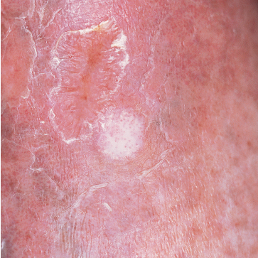
A 57-year-old man with a history of venous stasis leg ulceration wondered about the “white spots” on his leg. The condition is atrophie blanche, which manifests as smooth, ivory-white macules and plaques of sclerosis stippled with telangiectasia that often are surrounded by mild to moderate pigmentation.
- Polycythemia Vera

Four months after a patchy, macular, erythematous spot erupted on the dorsum of a 63-year-old woman’s left foot, the area became ulcerated, tender, and painful. The 1.2-cm ulcer was covered by a hemorrhagic crust surrounded by a cyanotic reticular discoloration of the skin.
- Nasal Hemangioma

A 21-year-old woman had experienced recurrent nosebleeds and pain in her nose for the previous 2 months. Physical examination revealed an extremely vascular, slowly enlarging intranasal growth on the anterior surface of the septum.
- Erythema Elevatum Diutinum

This rare condition affects both men and women. The average age at onset is 53 years. The lesions are deep brownish red to purple papules, nodules, and plaques. Blisters and ulcers also can occur, as pictured here.
- Schamberg Disease

These orange-to-brown macules with red puncta, or cayenne pepper spots, are typical of Schamberg disease (progressive pigmented purpuric dermatosis). The cause of this disorder is unknown, but it may be related to a cellular immune reaction or drug reaction.
A biopsy of tissue from the affected areas of a 70-year-old man’s legs revealed perivascular lymphohistiocytic infiltrate of the epidermis with focal red blood cell extravasation. These findings confirmed the diagnosis of Schamberg disease.
Vascular Disease
Slideshow: Skin Signs of Vascular Disorders
07/22/2021
