
Peer Reviewed
The Sag Sign: Posterior Cruciate Ligament Injury of the Knee
Authors:
Jonathan Ermer, BS; Apoorva Sharma, MS; and Shailendra Saxena, MD, PhD
Department of Family Medicine, Creighton University Medical Center, Omaha, Nebraska
Citation:
Ermer J, Sharma A, Saxena S. The sag sign: posterior cruciate ligament injury of the knee. Consultant. 2019;59(12):380-382.
A 55-year-old man presented with pain and instability in his left knee that had been worsening over the past month. He had no history of recent trauma but played football and baseball when he was younger. He reported feeling that his left knee would “give way,” especially when it was slightly bent. He rated the pain as 7 of 10 at its worst. He denied swelling on the knee and any fevers or chills. He also denied any feeling of the knee catching or getting stuck. He worked as a rehabilitation specialist and was on his feet most of the day, and the injury had been significantly impacting his ability to perform his job.
History. His medical history was significant for hypertriglyceridemia, hyperlipidemia, hypertension, and gout. His last acute attack of gout had occurred approximately 18 months ago. The gout had involved his left first metatarsophalangeal joint, and he had been given oral prednisone for any future acute gout attacks. His symptoms had cleared on their own, and he had not taken any prednisone. His current medications included fenofibrate, lisinopril, and simvastatin. He had been taking ibuprofen as needed for the knee pain, but it had been providing him less relief. He had never smoked, rarely drank alcohol, and did not use recreational drugs.
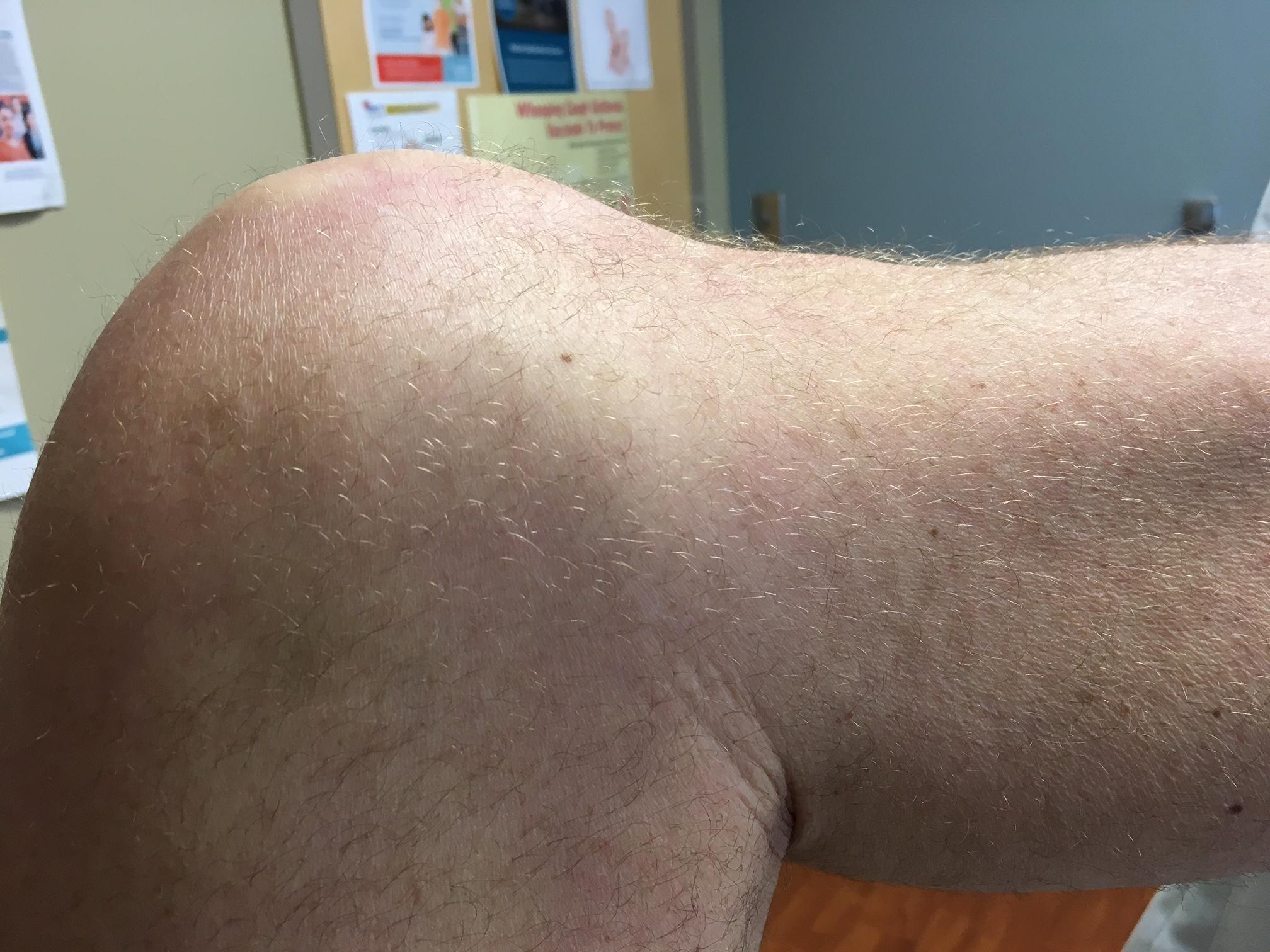
Physical examination. Vitals signs were all within normal limits except for a slightly elevated blood pressure of 138/78 mm Hg. The patient was overweight, with a body mass index of 27.8 kg/m2. On physical examination, the heart, lungs, and abdomen were normal. The right knee was normal. On examination of the left knee, no swelling was present. There was moderate tenderness on the left medial meniscus and bony tenderness on the left medial knee. The Lachman test was positive for both anterior and posterior movement, and the anterior and posterior drawer tests were both positive. The posterior sag sign was present on the left, suggestive of a posterior cruciate ligament (PCL) tear (Figure 1). There was no laxity of the knee during adduction or abduction. The fulcrum test was negative, making a stress fracture of the femur less likely. The McMurray test was negative for meniscal tear.

Diagnostic tests. Radiographs of the knee were ordered due to bony tenderness the findings were within normal limits. A magnetic resonance imaging (MRI) scan was ordered, the results of which showed a chronic complete tear of the PCL and a torn medial meniscus (Figure 2).

After the MRI, the results were discussed with the patient in clinic. The patient was referred to an orthopedic surgeon for an appointment in 2 weeks. He was prescribed meloxicam for his knee pain until he could be seen by the orthopedist. The patient was instructed to follow-up in 4 weeks.
Discussion. Understanding the anatomy of the knee joint is essential in detecting and diagnosing knee injuries. The knee joint consists of 3 articulations—two femorotibial and one femoropatellar. The joint is stabilized by a series of ligaments, a fibrous external joint capsule, and a large suprapatellar bursa. Extracapsular and intra-articular ligaments play an important role in strengthening the joint and enabling flexion and extension of the knee. Externally, the fibular collateral ligament and tibial collateral ligament attach to and strengthen the joint capsule.1
The structures within the knee joint include the anterior cruciate ligament (ACL), the PCL, and the medial and lateral menisci. The cruciate ligaments are so called because they form an X within the knee joint capsule. This point serves as a pivot for the rotational movements of the knee.1 Functionally, the ACL and PCL help prevent anterior and posterior displacement of the femur on the tibia. The PCL is the stronger of the two cruciate ligaments and as such is injured less often. The PCL arises from the posterior tibia, passes superiorly and anteriorly to the ACL, and attaches to the femur (Figure 3).2 During weight-bearing flexion, the PCL prevents anterior displacement of the femur on the tibia. Thus, the PCL is the main stabilizing factor for the femur.

In addition to the cruciate ligaments, the menisci of the knee joint are fibrocartilage plates that articulate with and deepen the surface of the tibia. They also aid in shock absorption and thus are commonly torn during high-impact injuries. The medial and lateral menisci are connected anteriorly via the transverse ligament of the knee.
PCL injuries are uncommon, accounting for approximately 0.65% of all athletic knee injuries in one large long-term study.3 PCL injuries can often be subtle and therefore more difficult to diagnose clinically.4,5 The history and physical examination becomes important in accurately diagnosing a PCL tear.
Several examination techniques and signs are particularly helpful in diagnosing PCL tears in the outpatient setting, such as the posterior drawer test, the Lachman test, and as in this patient’s case, the posterior sag sign, a lesser-known physical examination finding.
The posterior sag sign is a highly specific and sensitive sign for diagnosing PCL injuries, with values of 83% and 100%, respectively.6 PCL injuries usually occur when the knee is flexed or during head-on collisions of the tibia, such as when the tibia strikes the dashboard during a motor vehicle accident. A torn PCL allows the free tibia to slide posteriorly under the fixed femur, creating the posterior sag sign. A positive sag sign is characteristic of PCL injuries and results when gravity pulls the tibia downward greater than 10 mm. Clinicians can also use the posterior drawer test to evaluate the injury. A positive drawer sign occurs if the patient’s tibia slides posteriorly. Physical examination of the patient in this case revealed a positive sag sign and a positive posterior drawer sign.
PCL tears often include injuries to other structures of the knee. For example, injury to the lateral side of the extended knee or excessive lateral rotation can cause the medial meniscus to detach from the joint capsule.1 The McMurray test can be used to determine the presence of a meniscal tear.7 Generally, meniscal tears involve the medial meniscus since it is immobile. Pain upon medial rotation of the tibia on the femur is indicative of medial meniscus injury. An MRI scan of our patient revealed both a torn PCL and a torn medial meniscus.
In general, the location and severity of the PCL tear is important in determining whether PCL repair will be beneficial to the patient. Tears that occur at nonattachment sites are referred to as midsubstance tears and are more common than tears at the origin or insertion of the ligament.8 Operative reattachment using sutures is the treatment of choice for midsubstance PCL injuries.9 However, nonoperative management is recommended for isolated grade I or II PCL tears.10 Additionally, data suggest that repair or partial excision of the medial meniscus can also help patients regain stability, but whether this intervention is appropriate must be evaluated on a case-by-case basis.
References:
- Moore KL, Dalley AF, Agur AMR. Knee joint. In: Moore KL, Dalley AF, Agur AMR. Clinically Oriented Anatomy. 7th ed. Philadelphia, PA: Wolters Kluwer/Lippincott Williams & Wilkins; 2014:634-644.
- Van Dommelen BA, Fowler PJ. Anatomy of the posterior cruciate ligament: a review. Am J Sports Med. 1989;17(1):24-29.
- Majewski M, Susanne H, Klaus S. Epidemiology of athletic knee injuries: a 10-year study. Knee. 2006;13(3):184-188.
- McAllister DR, Petrigliano FA. Diagnosis and treatment of posterior cruciate ligament injuries. Curr Sports Med Rep. 2007;6(5):293-299.
- Wahl CJ, Chin PC. Surgical treatment of combined PCL/lateral-sided injuries. In: Fanelli GC, ed. Posterior Cruciate Ligament Injuries: A Practical Guide to Management. Cham, Switzerland: Springer International Publishing; 2015:189-208.
- Malanga GA, Andrus S, Nadler SF, McLean J. Physical examination of the knee: a review of the original test description and scientific validity of common orthopedic tests. Arch Phys Med Rehabil. 2003;84(4):592-603.
- McMurrays test. Physiopedia. https://www.physio-pedia.com/McMurrays_Test. Revised August 29, 2018. Accessed February 12, 2019.
- Kongcharoensombat W, Nakamae A, Adachi N, et al. Mid-substance tear of the anterior and posterior cruciate ligaments in children: a report of three patients. Knee Surg Sports Traumatol Arthrosc. 2009;17(8):964-967.
- Barrett GR, Savoie FH. Operative management of acute PCL injuries with associated pathology: long-term results. Orthopedics. 1991;14(6):687-692.
- Montgomery SR, Johnson JS, McAllister DR, Petrigliano FA. Surgical management of PCL injuries: indications, techniques, and outcomes. Curr Rev Musculoskel Med. 2013;6(2):115-123.