Peer Reviewed
Right Lower Eyelid and Cheek Tenderness and Swelling
Correct answer: C. Biopsy
Discussion. The differential diagnosis of recurrent facial swelling is broad and includes infectious causes such as erysipelas, underlying dental abscess, or herpetic infection, as well as allergic hypersensitivity reactions. In our case, the absence of fever, drainage, vesicles on clinical inspection, or abnormalities on CT scan indicated that a diagnostic biopsy was warranted.
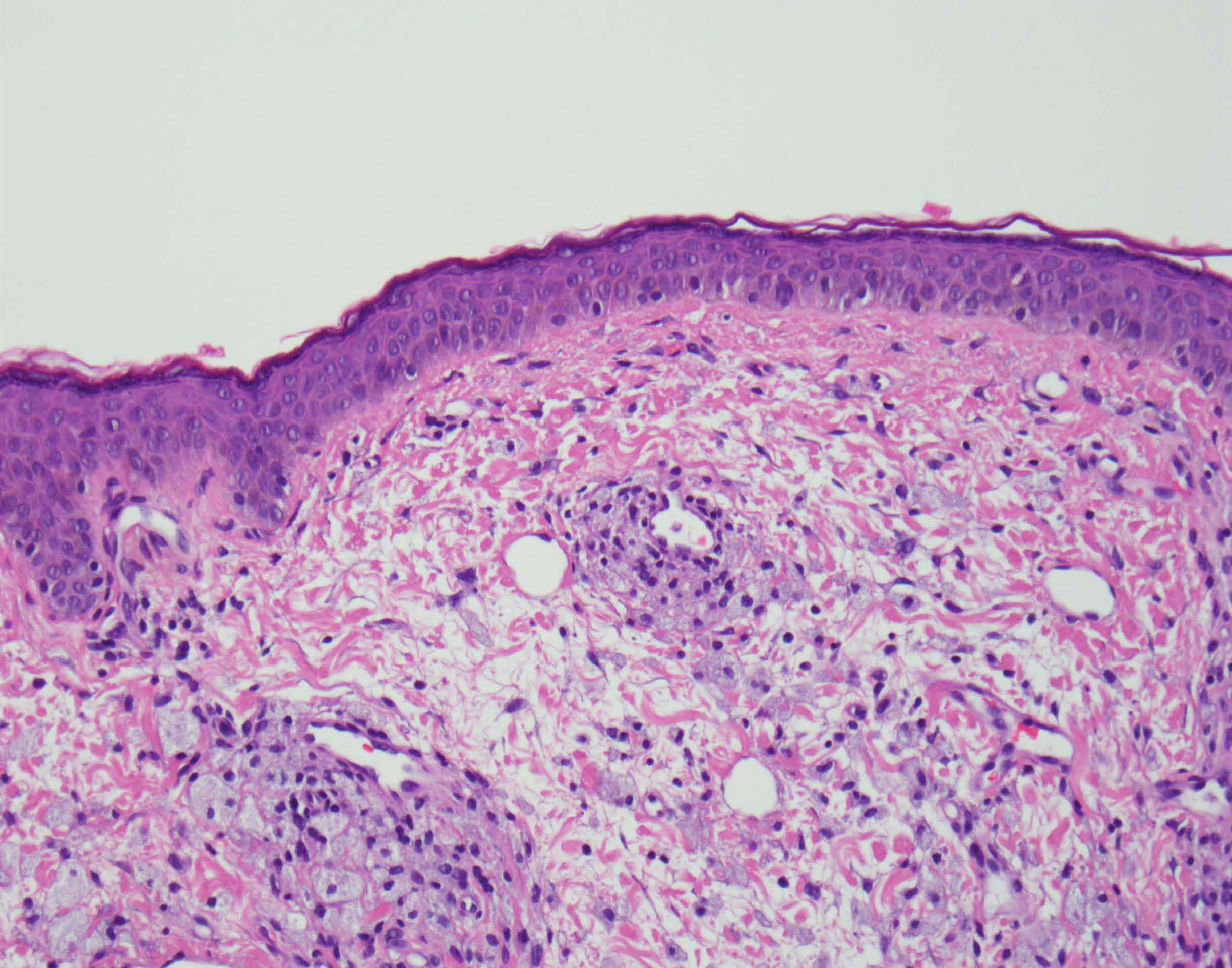
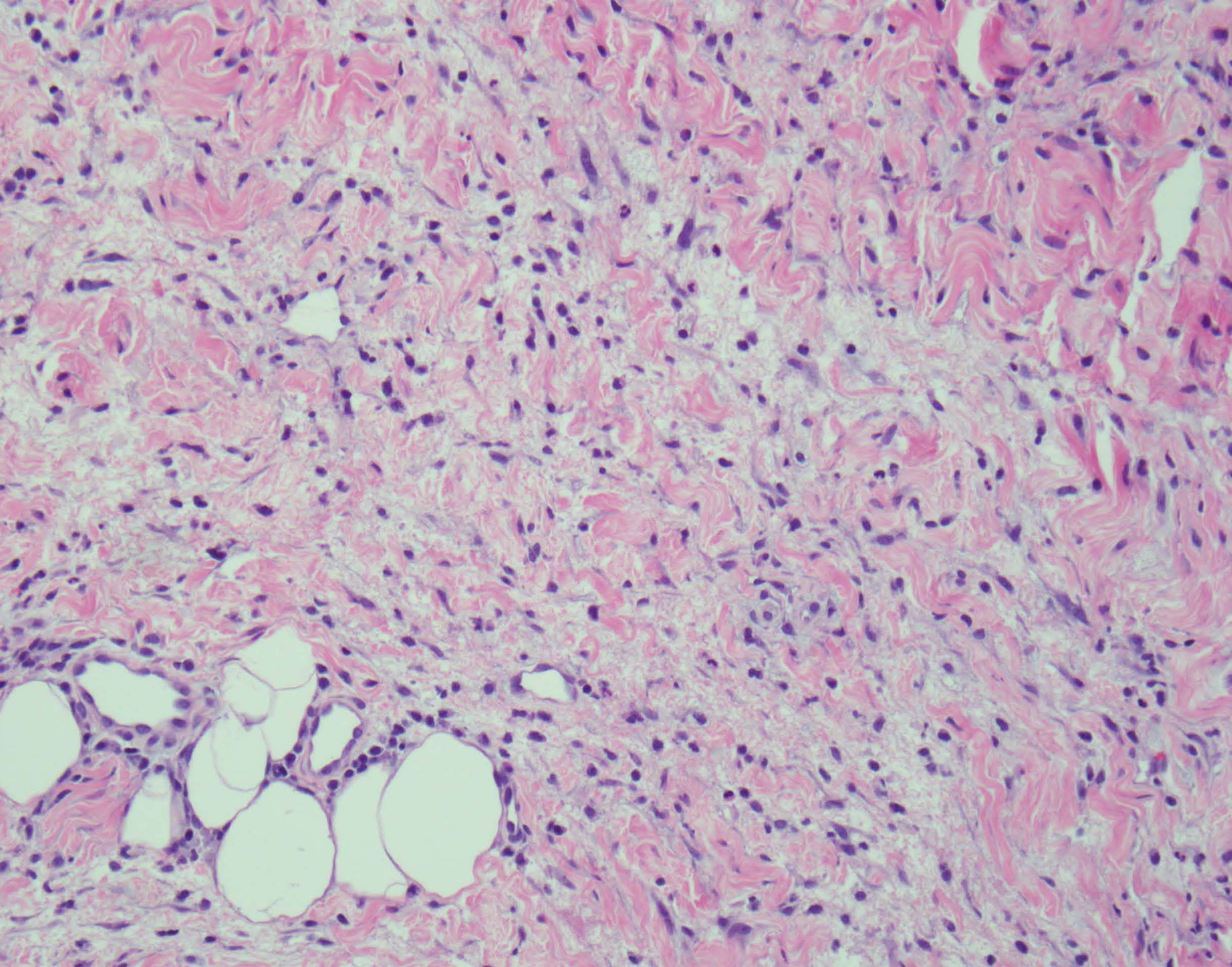
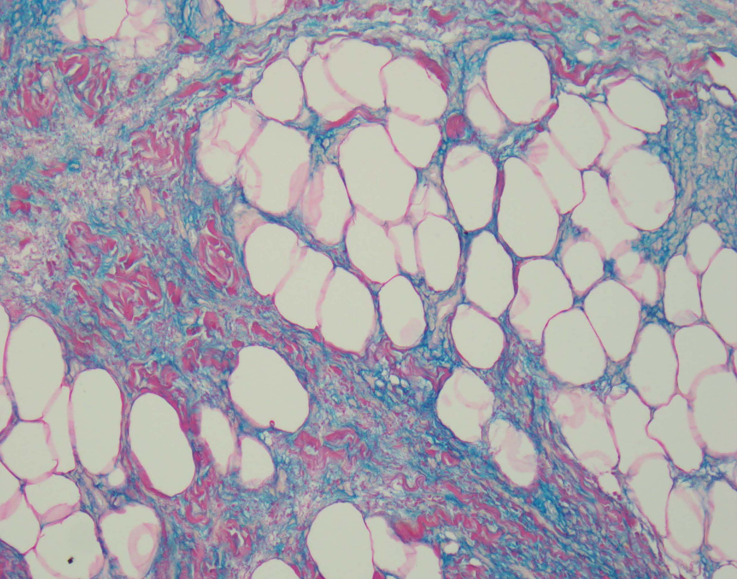
Upon biopsy, histologic examination revealed foam cells around small blood vessels and adnexal structures (Figure 2). Chronic inflammation in the dermis and amorphous material (Figure 3) were present. Colloidal iron stain highlighted amorphous material in the dermis (Figure 4). The collections of foam cells detected in the patient's lower eyelid area are characteristic of the xanthoma-like reaction to hyaluronic acid (HA) filler. Along with the patient's clinical appearance and course, her condition was diagnosed as xanthomatous filler reaction associated with delayed hypersensitivity. Other adverse local reactions may include bruising, redness, granuloma formation, and infection.

Figure 2. Numerous foam cells are noted on a perivascular location. A patchy lymphohistiocytic infiltrate is encountered (hematoxylin and eosin-stained sections; original magnification 200×).
Figure 3. Amorphous material is noted in the dermis along with chronic inflammation (hematoxylin and eosin-stained sections; original magnification 200 ×).
Figure 4. Colloidal iron stain highlights the amorphous material, supporting the histologic impression of mucin.
Injection of HA soft tissue fillers is an increasingly popular technique to enhance facial appearance because of their safety, effectiveness, and ease of use.1 As more individuals undergo this type of cosmetic treatment, clinicians may encounter patients with unexpected reactions to these fillers. Injected HA can migrate and lead to delayed reactions, which can be more difficult to diagnose.2 Foreign body granulomatous reaction is a rare, delayed reaction to HA filler, which is typically associated with poor injection technique.3. Lesions may be erythematous, and some may have a yellow hue. Hyaluronic acid fillers can lead to lipid deposition that mimics xanthelasma palpeprum.4-7
The immunologic reactions underlying delayed hypersensitive reactions to HA fillers are not well understood. Delayed inflammatory reactions have been reported to occur within hours to weeks following infection with or vaccination against SARS-CoV-2.8-10 Inflammatory reactions may include angioedema, lip swelling, and localized facial swelling.9 Reactions have been noted to occur days to weeks after vaccination.11
Treatment options that have been used with varying degrees of success include hyaluronidase injection, intralesional corticosteroids, excision, and laser ablation. Lisinopril has been reported to help with delayed reactions after SARS CoV-2 infection.9 Granuloma formation can sometimes be ameliorated by a hyaluronidase injection.2
Our patient was treated with intralesional hyaluronidase, intralesional corticosteroids, oral steroids, topical steroids, and several oral antibiotics. Her lesions have gradually resolved over months.
Our case highlights that as HA fillers become more popular, clinicians should be aware of the possibility of a hypersensitivity reaction so that early diagnosis can lead to prompt treatment.
References
1. Mandal P, Gama F. The use of periocular fillers in aesthetic medicine. J Plast Reconstr Aesthet Surg. 2021;74(7):1602-1609. doi:10.1016/j.bjps.2020.12.079
2. Chang JR, Baharestani S, Salek SS, Piluek WJ, Eberhart CG, McCulley TJ. Delayed superficial migration of retained hyaluronic acid years following periocular injection. Ophthalmic Plast Reconstr Surg. 2017;33(3S suppl 1):S116-S118. doi:10.1097/IOP.0000000000000434
3. Caldas Pozuelo C, Domínguez De Dios J, Mota Rojas X. Multiple oral granulomatous nodules to hyaluronic acid filler. J Cosmet Dermatol. 2020;19(12):3453-3455. doi:10.1111/jocd.13734
4. Or L, Eviatar JÁ, Massry GG, Bernardini lFP, Harstein ME. Xanthelasma-like reaction to filler injection. Ophthal Plast Reconstr Surg. 2017;33(4):244-247. doi:10.1097/IOP.0000000000000722
5. Liu A, Kollipara R, Hoss E, Goldman MP. Lower eyelid xanthelasma following hyaluronic acid filler injections to the tear troughs. J Cosmet Dermatol. 2021;20(10):3190-3192. doi:10.1111/jocd.14166
6. D'Acunto C, Pazzaglia M, Raone B, et al. Xanthelasma palpebrarum: a new adverse reaction to intradermal fillers? Br J Dermatol. 2013;168(2):437-439. doi:10.1111/j.1365-2133.2012.11152.x
7. Seike M, Ikeda M, Matsumoto M, Hamada R, Takeya M, Kodama H. Hyaluronan forms complexes with low density lipoprotein while also inducing foam cell infiltration in the dermis. J Dermatol Sci. 2006;41(3):197-204. doi:10.1016/j.jdermsci.2005.10.008
8. Michon A. Hyaluronic acid soft tissue filler delayed inflammatory reaction following COVID-19 vaccination - A case report. J Cosmet Dermatol. 2021;20(9):2684-2690. doi:10.1111/jocd.14312
9. Bachour Y, Bekkenk MW, Rustemeyer T, Kadouch JA. Late inflammatory reactions in patients with soft tissue fillers after SARS-CoV-2 infection and vaccination: A systematic review of the literature. J Cosmet Dermatol. Published online February 12, 2022. doi:10.1111/jocd.14840
10. Arron ST, Neuhaus IM. Persistent delayed-type hypersensitivity reaction to injectable non-animal-stabilized hyaluronic acid. J Cosmet Dermatol. 2007;6(3):167-171. doi:10.1111/j.1473-2165.2007.00331.x
11. Beamish IV, Bogoch II, Carr D. Delayed inflammatory reaction to dermal fillers after COVID-19 vaccination: a case report. CJEM. 2022;24(4):444-446. doi:10.1007/s43678-022-00289-x