
Peer Reviewed
A Case of Infantile Hemangioma
AUTHORS:
Alexander K. C. Leung, MD1,2 • Joseph M. Lam, MD3 • Kin Fon Leong, MD4
AFFILIATIONS:
1Department of Pediatrics, University of Calgary, Calgary, Alberta, Canada
2Alberta Children’s Hospital, Calgary, Alberta, Canada
3Department of Pediatrics and Department of Dermatology and Skin Sciences, University of British Columbia, Vancouver, British Columbia, Canada
4Pediatric Institute, Kuala Lumpur General Hospital, Kuala Lumpur, Malaysia
CITATION:
Leung AKC, Lam JM, Leong KF. A case of infantile hemangioma. Consultant. 2021;61(11):e8-e11. doi:10.25270/con.2020.10.00005
DISCLOSURES:
The authors report no relevant financial relationships.
CORRESPONDENCE:
Alexander K. C. Leung, MD, #200, 233 16th Ave NW, Calgary, AB T2M 0H5, Canada (aleung@ucalgary.ca)
A healthy 5-month-old girl presented with a vascular plaque over the back that had been noted a few weeks after birth. The plaque had begun as an erythematous patch that had grown fairly rapidly over the first 2 months, with slower growth recently. She had been born to a gravida 3, para 2, 30-year-old mother at term following an uncomplicated pregnancy and normal spontaneous delivery. Her Apgar scores were 7 and 9 at 1 minute and 5 minutes, respectively. Her birth weight was 2.74 kg, length was 49 cm, and head circumference was 39.2 cm. She had been exclusively breastfed and was thriving.
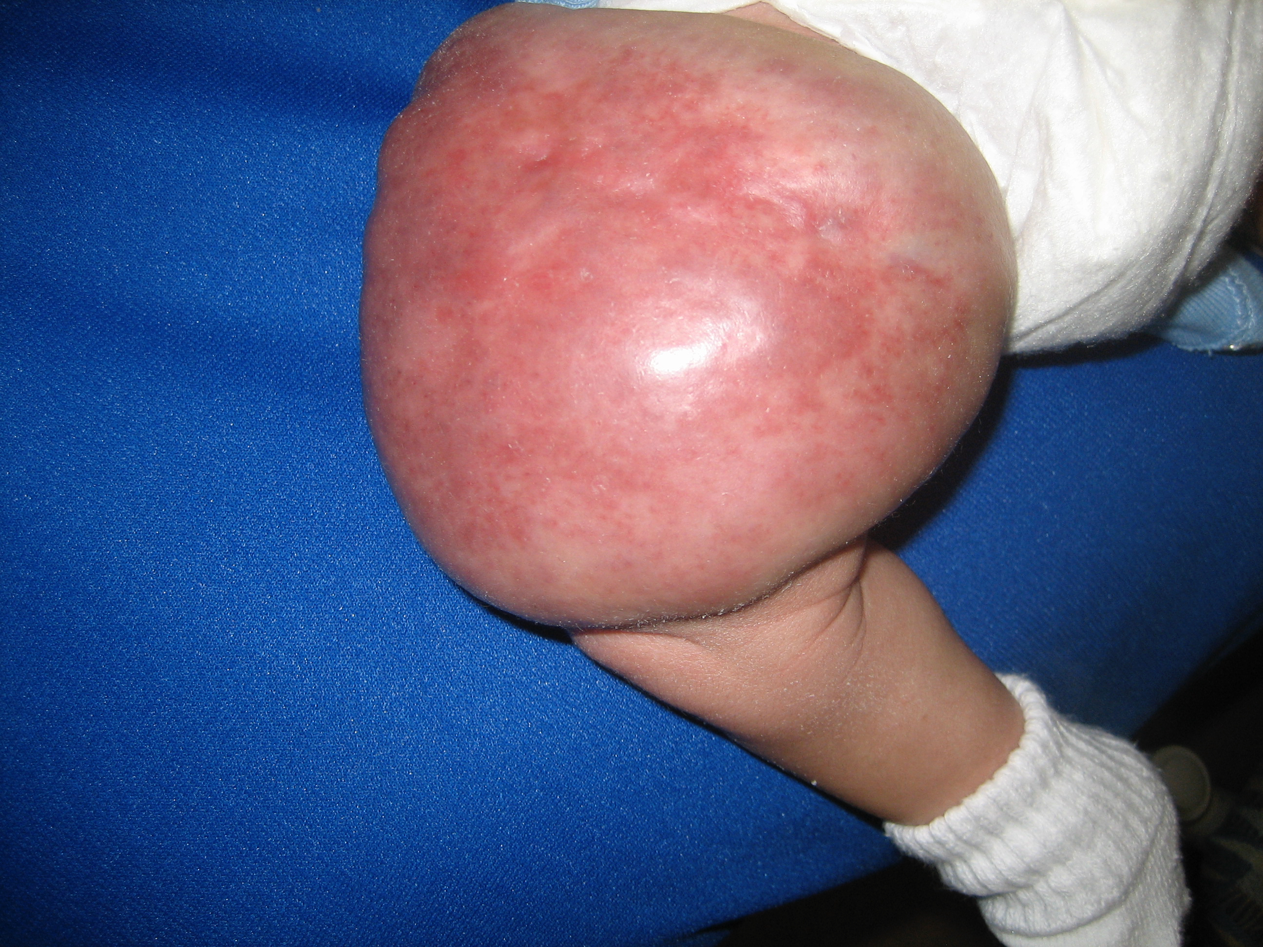
Physical examination revealed a happy-appearing girl in no distress. Her vital signs were normal. A large vascular plaque was noted over the left mid back (Figure 1). The rest of the physical examination findings were normal. In particular, she did not have apparent visceromegaly or dysmorphic features.

Based on the timing of appearance of the lesion, its rapid growth in the first few months of life, its slower growth later on, and the color of the lesion, a diagnosis of infantile hemangioma was made. The parents were reassured of the benign nature of the lesion and that it would resolve spontaneously with time.
DISCUSSION
Typically, an infantile hemangioma (formerly referred to as “capillary hemangioma” or “strawberry hemangioma”) presents as an asymptomatic, sharply demarcated, bright red, protuberant plaque or nodule with an either bosselated or smooth surface. Infantile hemangiomas are the most common vascular tumors of infancy, accounting for approximately 55% of cases.1,2
Infantile hemangiomas typically appear in the first few weeks of life as areas of pallor, followed by telangiectatic or faint red macules or patches with surrounding pallor.1-5 Characteristically, infantile hemangiomas grow rapidly in the first 3 to 6 months of life (proliferative phase), beyond the growth rate of the infant, as illustrated in the presented case.6 The growth rate then slows to parallel the growth of the child (quiescent or plateau phase).1,2,6 Involution typically begins by the time the child is 1 year old (involution phase). Approximately 50% of infantile hemangiomas have involuted by age 5, 70% by age 7, 90% by age 9, and 95% by age 12.1,2,7 Although infantile hemangiomas can appear anywhere on the skin, they have a predilection for the head and neck region.2,8-10
The differential diagnosis includes, among others, congenital hemangiomas, port-wine stain, salmon patch, and pyogenic granuloma. The characteristic features of these conditions are summarized in the Table. Congenital hemangiomas are benign high-flow vascular tumors that proliferate in utero and are fully developed at birth and do not proliferate further postnatally.11 Typically, a congenital hemangioma presents as a solitary bluish or violaceous nodule or, less commonly, as a plaque with overlying telangiectases and venules.11 A pale surrounding halo is quite common. A central ulcer may also occur. Sites of predilection include the head, neck, and limbs, especially around the elbows and knees.12 Two major clinical subtypes are recognized, namely, rapidly involuting congenital hemangiomas (RICH) and noninvoluting congenital hemangiomas (NICH), based on their clinical progression. RICHs typically shrink rapidly after birth with complete involution usually by 6 to 14 months of age (Figure 2).11,13,14 NICHs, on the other hand, do not regress but instead grow proportionally with the child and remain static throughout the lifetime (Figure 3).2,11,13 A subset of congenital hemangiomas, known as partially involuting congenital hemangiomas (PICHs), begin with a phase of involution lasting until 12 to 30 months of age, at which time the involution halts.2,11,15,16


Figure 2. A rapidly involuting congenital hemangioma on the right knee of an infant at 3 days of age (A, left) and again at 6 months of age (B, right).
Typically, a port-wine stain, also known as nevus flammeus, presents as a sharply demarcated pink or red macule or patch at birth (Figure 4).17 Port-wine stains may appear to fade during the first 12 months of life due to the natural fall in hemoglobin. The capillaries become more ectatic with age, and the color thereafter gradually deepens. Port-wine stains often become dark red during adolescence and violaceous with advancing age.17 Although they are initially macular, the surface might become irregular, thickened and nodular over time.17 Port-wine stains can occur in any cutaneous location, but facial involvement is most common. They are usually unilateral, segmental, and do not follow the lines of Blaschko. These lesions tend to persist throughout life. Although usually an isolated finding, port-wine stains can be a cutaneous feature of Sturge-Weber syndrome and Klippel-Trenaunay syndrome.

A salmon patch, also known as nevus flammeus simplex, typically presents as a scarlet to pink macule that is blanchable and that deepens in color with vigorous activity, crying, straining with defecation, breath-holding, or changes in ambient temperature (Figure 5).18 They are usually bright red or pink in White infants and are darker in Asian or Black infants. The lesions are most commonly found on the nape, followed by the glabella and eyelids. Salmon patches are usually symmetric, with lesions on both eyelids and/or on both sides of the midline. Prominent lesions in the glabella may be associated with Beckwith-Wiedemann syndrome and fetal alcohol syndrome.18 Despite their midline location, most salmon patches, except those in the sacral area, are not associated with spinal dysraphism.18 Salmon patches on the eyelids and glabella usually disappear by 2 to 3 years of age.19 Nuchal and sacral lesions tend to persist longer.19

Pyogenic granuloma, also known as granuloma pyogenicum or lobular capillary hemangioma, is an acquired, benign vascular proliferation of the skin and mucous membranes.20 Cutaneous pyogenic granulomas are commonly located on the head and neck (62.5%), trunk (19.7%), and extremities (17.9%), especially the fingers.21 In the oral cavity, pyogenic granulomas are more frequent on the gingiva. The condition is more prevalent in children, adolescents, and pregnant women, and has been associated with minor trauma, ingrown nails, and medications. Pyogenic granuloma typically develops as a small erythematous papule on the skin or oral mucosal surface. The papule usually enlarges quickly to a few millimeters and occasionally a few centimeters over weeks, and growth stabilizes over several months. Clinically, pyogenic granuloma presents as a soft, dome-shaped papule with a smooth, glistening, erosive, or friable surface (Figure 6). The color is usually bright red to dusky red initially. With time, the vascularity decreases, and the lesion tends to become more collagenized and pink. Characteristically, the lesion is painless. Due to its pronounced vascularity, a pyogenic granuloma tends to bleed and ulcerate even with very minor trauma. As such, patients often present with the “Band-Aid sign,” where the patient presents with the lesion covered by an adhesive bandage. The lesion is usually solitary, but multiple lesions may occur. Pyogenic granulomas that develop during pregnancy tend to resolve on their own after delivery. Spontaneous resolution of other pyogenic granulomas is rare, and they are usually removed surgically or treated with topical imiquimod or topical β-blockers.

REFERENCES:
- Leung AKC, Barankin B, Hon KL. Infantile hemangioma. Pediatr Neonatal Nurs Open J. 2014;1(1):6-11. doi:10.17140/PNNOJ-1-102
- Leung AKC, Lam JM, Leong KF, Hon KL. Infantile hemangioma: an updated review. Curr Pediatr Rev. Published online May 7, 2020. doi:10.2174/1573396316666200508100038
- Adams DM, Ricci KW. Infantile hemangiomas in the head and neck region. Otolaryngol Clin North Am. 2018;51(1):77-87. doi:10.1016/j.otc.2017.09.009
- Bilodi AKS, Singh S, Ebenezer DA, Suman P, M R. Capillary haemangioma of the right elbow and forearm in new born child. J Clin Diagn Res. 2013;7(12):2941-2942. doi:10.7860/JCDR/2013/6379.3797
- Chisti M, Banka N, Alfadley A. Pallor sign: an indicator of hemangioma in evolution. J Cutan Med Surg. 2012;16(6):451-452. doi:10.1177/120347541201600619
- Beck DO, Gosain AK. The presentation and management of hemangiomas. Plast Reconstr Surg. 2009;123(6):181e-91e. doi:10.1097/PRS.0b013e3181a65c59
- Püttgen KB. Diagnosis and management of infantile hemangiomas. Pediatr Clin North Am. 2014;61(2):383-402. doi:10.1016/j.pcl.2013.11.010
- Atherton DJ. Infantile haemangiomas. Early Hum Dev. 2006;82(12):789-795. doi:10.1016/j.earlhumdev.2006.09.011
- Holland KE, Drolet BA. Approach to the patient with an infantile hemangioma. Dermatol Clin. 2013;31(2):289-301. doi:10.1016/j.det.2012.12.006
- Soliman YS, Khachemoune A. Infantile hemangiomas: our current understanding and treatment options. Dermatol Online J. 2018;24(9):13030/qt5jt8q9km.
- Leung AKC, Leong KF, Barankin B. A male infant born with an ulcerated vascular mass. Paediatr Child Health. 2019;24(1):7-9. doi:10.1093/pch/pxy071
- Frieden IJ, Rogers M, Garzon MC. Conditions masquerading as infantile haemangioma: part 1. Australas J Dermatol. 2009;50(2):77-98. doi:10.1111/j.1440-0960.2009.00514_1.x
- Harter N, Mancini AJ. Diagnosis and management of infantile hemangiomas in the neonate. Pediatr Clin North Am. 2019;66(2):437-459. doi:10.1016/j.pcl.2018.12.011
- Hon KLE, Burd A, Chu WC, Lee V, Li CK. Propranolol for infantile hemangiomas: strawberry matters? Indian J Pediatr. 2012;79(1):130-131. doi:10.1007/s12098-011-0527-5
- Ayturk UM, Couto JA, Hann S, et al. Somatic activating mutations in GNAQ and GNA11 are associated with congenital hemangioma. Am J Hum Genet. 2016;98(6):1271. doi:10.1016/j.ajhg.2016.05.010
- Boull C, Maguiness SM. Congenital hemangiomas. Semin Cutan Med Surg. 2016;35(3):124-127. doi:10.12788/j.sder.2016.045
- Leung AKC. Port-wine stain associated with maternal use of lithium carbonate. J Natl Med Assoc. 1987;79(8):877-878.
- Leung AKC, Telmesani AM. Salmon patches in Caucasian children. Pediatr Dermatol. 1989;6(3):185-187. doi:10.1111/j.1525-1470.1989.tb00813.x
- Leung AKC, Barankin B, Hon KL. Persistent salmon patch on the forehead and glabellum in a Chinese adult. Case Rep Med. 2014;2014:139174. doi:10.1155/2014/139174
- Garcia-Monaco RD, Giachetti A. Infantile hemangioma or kaposiform hemangioendothelioma? J Vasc Interv Radiol. 2014;25(5):810. doi:10.1016/j.jvir.2014.01.024
- Lin RL, Janniger CK. Pyogenic granuloma. Cutis. 2004;74(4):229-233.