
Peer Reviewed
An Atlas of Lumps and Bumps: Part 12
AUTHORS:
Alexander K. C. Leung, MD1,2—Series Editor • Benjamin Barankin, MD3 • Joseph M. Lam, MD4 • Andrew A. H. Leung, BSc5
AFFILIATIONS:
1Department of Pediatrics, University of Calgary, Calgary, Alberta, Canada
2Alberta Children’s Hospital, Calgary, Alberta, Canada
3Toronto Dermatology Centre, Toronto, Ontario, Canada
4Department of Pediatrics and Department of Dermatology and Skin Sciences, University of British Columbia, Vancouver, British Columbia, Canada
5Faculty of Medicine, St. George’s University, Grenada
CITATION:
Leung AKC, Barankin B, Lam JM, Leung AAH. An atlas of lumps and bumps, part 12. Consultant. 2022;62(1):e15-e16. doi:10.25270/con.2021.12.00007
DISCLOSURES:
Dr Leung is the series editor. He was not involved with the handling of this paper, which was sent out for independent external peer review.
CORRESPONDENCE:
Alexander K. C. Leung, MD, #200, 233 16th Ave NW, Calgary, AB T2M 0H5, Canada (aleung@ucalgary.ca)
EDITOR’S NOTE:
This article is part of a series describing and differentiating dermatologic lumps and bumps. To access previously published articles in the series, visit https://bit.ly/35J1I1v.
Bohn Nodule
A Bohn nodule is characterized by a firm, whitish lesion on the buccal and, less commonly, the lingual aspect of the maxillary or the mandibular alveolar ridge (Figures 1 and 2).1-3 The lesion has a rice-like appearance and can be solitary or multiple. Once thought of as a mucous gland cyst, recent studies suggest that it is either a keratin cyst derived from remnants of odontogenic epithelium over the dental lamina or remnants of minor salivary glands.2,3 The condition is asymptomatic and does not interfere with feeding.1 The nodules are named after Heinrich Bohn, a German pediatrician who first described them in 1866. A Bohn nodule is often mistaken by parents as a natal or erupting tooth.4 Other differential diagnoses include Epstein pearl, congenital epulis, pyogenic granuloma, and dental lamina cyst (gingival cyst). Bohn nodules occur more frequently in full-term vs preterm infants.5,6 In one study, folic acid consumption during pregnancy was significantly associated with Bohn nodules (odds ratio: 1.79; 95% CI: 1.23-2.55).5 The lesions are self-limiting and usually disappear by age 3 months.


Epstein Pearls
Clinically, Epstein pearls present as pearly white or whitish-yellow papules on the roof of the mouth (Figure 3). The condition was first described by Alois Epstein, a Prague pediatrician, in 1880 and now bears his name.7,8 Presumably, Epstein pearls are caused by epithelium trapped between the palatal shelves during palatal fusion.8 Some authors suggest that Epstein pearls may result from remnants of the minor salivary glands of the palate.8 Histologically, Epstein pearls are keratin-containing cysts lined by stratified squamous epithelium.8 Epstein pearls are seen in 60% to 90% of neonates.9-12 The condition is more common in individuals with Japanese race, followed by White and Black race.11 The sex ratio is approximately equal.7 Epstein pearls are more prevalent in infants born to multigravida mothers.7,9 Data corelating the length of gestation and prevalence of Epstein pearls are conflicting.5,9,13

Typically, Epstein pearls are seen in the mid-palatal raphe at the junction of the hard and soft palate.7,11 In infants with cleft palate, the lesions are located at the margins of the palatal shelves.7,11 Lesions can be solitary or, more commonly, multiple and tend to cluster. The lesion varies from 1 mm to 3 mm in diameter. The size of the lesion usually does not progress over time.7 On palpation, larger lesions can be appreciated when the infant is sucking on the examiner’s finger. Epstein pearls are asymptomatic and do not cause problems with feeding.
Similar to Bohn nodules, Epstein pearls usually resolve spontaneously by age 3 months.11 As such, treatment is not necessary.
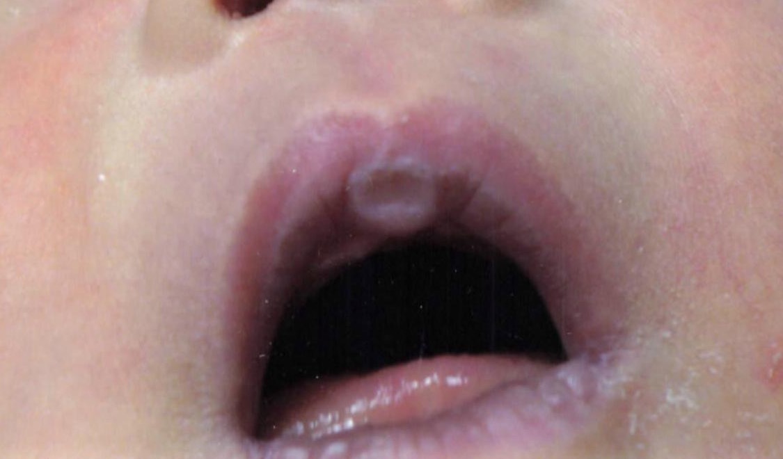
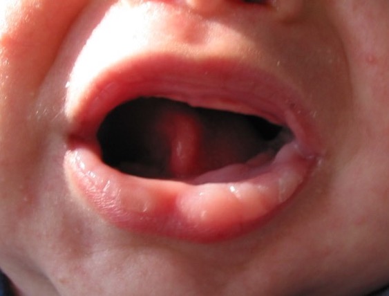
Neonatal Sucking Pads of the Lips
Sucking pads can develop on the lips in the postnatal period as a result of sucking on a nipple or pacifier.14 The pads are hyperkeratotic lesions and usually occur in the central portion of the lips (Figures 4-6).15 Sucking pads are caused by the combined intracellular edema and epithelial hyperplasia.15,16 They often persist for a few weeks to months and are of no medical significance.

Neonatal sucking pads should be distinguished from neonatal sucking blisters, which result from vigorous sucking by the infant during fetal life.16,17 Neonatal sucking blisters are always present at birth and are mainly located on the distal upper extremities.16,17 The lesions of neonatal sucking blisters usually resolve in days to weeks without treatment.18
1. Cambiaghi S, Gelmetti C. Bohn's nodules. Int J Dermatol. 2005;44(9):753-754. https://doi.org/10.1111/j.1365-4632.2005.02525.x
2. Dutta A, Ghosh SK, Nag SS. Bohn's nodules. Indian Pediatr. 2014;51(10):849-850.
3. Gupta N, Ramji S. Bohn's nodules: an under-recognised entity. Arch Dis Child Fetal Neonatal Ed. 2013;98(5):F464. https://doi.org/10.1136/archdischild-2012-302922
4. Cizmeci MN, Kanburoglu MK, Kara S, Tatli MM. Bohn's nodules: peculiar neonatal intraoral lesions mistaken for natal teeth. Eur J Pediatr. 2014;173(3):403. https://doi.org/10.1007/s00431-013-2173-6
5. Perez-Aguirre B, Soto-Barreras U, Loyola-Rodriguez JP, et al. Oral findings and its association with prenatal and perinatal factors in newborns. Korean J Pediatr. 2018;61(9):279-284. https://doi.org/10.3345/kjp.2017.06177
6. Zen I, Soares M, Sakuma R, Inagaki LT, Pinto LMCP, Dezan-Garbelini CC. Identification of oral cavity abnormalities in pre-term and full-term newborns: a cross-sectional and comparative study. Eur Arch Paediatr Dent. 2020;21(5):581-586. https://doi.org/10.1007/s40368-019-00499-5
7. Diaz de Ortiz LE, Mendez MD. Epstein Pearls. In: StatPearls. StatPearls Publishing; July 22, 2021. http://www.ncbi.nlm.nih.gov/books/nbk493177/
8. Epstein A. Über Epithelperlen in der. Mundhöhle neugeborener Kinder. Z Heilkd. 1880;1:59.
9. Gupta P, Faridi MM, Batra M. Physiological skin manifestations in twins: association with maternal and neonatal factors. Pediatr Dermatol. 2011;28(4):387-392. https://doi.org/10.1111/j.1525-1470.2011.01434.x
10. Haveri FT, Inamadar AC. A cross-sectional prospective study of cutaneous lesions in newborn. ISRN Dermatol. 2014;2014:360590. https://doi.org/10.1155/2014/360590
11. Richard BM, Qiu CX, Ferguson MW. Neonatal palatal cysts and their morphology in cleft lip and palate. Br J Plast Surg. 2000;53(7):555-558. https://doi.org/10.1054/bjps.2000.3410
12. Sachdeva M, Kaur S, Nagpal M, Dewan SP. Cutaneous lesions in new born. Indian J Dermatol Venereol Leprol. 2002;68(6):334-337.
13. Valdelice Cruz P, Bendo CB, Perez Occhi-Alexandre IG, Martins Paiva S, Pordeus IA, Castro Martins C. Prevalence of oral inclusion cysts in a Brazilian neonatal population. J Dent Child (Chic). 2020;87(2):90-97.
14. Aydin M, Hakan N, Zenciroglu A, Demirol HA. A rare location of sucking blister in newborn: the lips. Eur J Pediatr. 2013;172(10):1423-1424. https://doi.org/10.1007/s00431-013-2055-y
15. Heyl T, Raubenheimer EJ. Sucking pads (sucking calluses) of the lips in neonates: a manifestation of transient leukoedema. Pediatr Dermatol. 1987;4(2):123-128. https://doi.org/10.1111/j.1525-1470.1987.tb00765.x
16. Afsar FS, Cun S, Seremet S. Neonatal sucking blister. Dermatol Online J. 2019;25(11):13030/qt33b1w59j.
17. Monteagudo B, León-Muiños E. Neonatal sucking blisters. Indian Pediatr. 2010;47(9):794.