
AUTHORS:
Alexander K. C. Leung, MD1,2 —Series Editor • Benjamin Barankin, MD3 • Joseph M. Lam, MD4 • Kin Fon Leong, MD5
AFFILIATIONS:
1Department of Pediatrics, University of Calgary, Calgary, Alberta, Canada
2Alberta Children’s Hospital, Calgary, Alberta, Canada
3Toronto Dermatology Centre, Toronto, Ontario, Canada
4Department of Pediatrics and Department of Dermatology and Skin Sciences, University of British Columbia, Vancouver, British Columbia, Canada
5Pediatric Institute, Kuala Lumpur General Hospital, Kuala Lumpur, Malaysia
CITATION:
Leung AKC, Barankin B, Lam JM, Leong KF. An atlas of lumps and bumps, part 11. Consultant. 2021;61(21):e21-e23. doi:10.25270/con.2021.11.00006
DISCLOSURES:
Dr Leung is the series editor. He was not involved with the handling of this paper, which was sent out for independent external peer review.
CORRESPONDENCE:
Alexander K. C. Leung, MD, #200, 233 16th Ave NW, Calgary, AB T2M 0H5, Canada (aleung@ucalgary.ca)
EDITOR’S NOTE:
This article is part of a series describing and differentiating dermatologic lumps and bumps. To access previously published articles in the series, visit https://bit.ly/35J1I1v.
Pilomatricoma
A pilomatricoma, also known as pilomatrixoma or calcifying epithelioma of Malherbe, is a benign adnexal subcutaneous tumor derived from primitive epidermal germ cells differentiating toward hair matrix cells.1 Pilomatricomas account for approximately 1% of all benign skin nodules/cysts in childhood.2 The peak age of onset is in the first 2 decades of life and again between age 50 and 65 years.1-5 The female to male ratio is approximately 2:1.2 The condition is more common in White individuals than Asian individuals.6 Pilomatricomas can be familial.2 Activating mutations in β-catenin have been identified in approximately 75% of patients with pilomatricomas.7 The locus of this tumor has been mapped to the CTNNB1 gene on 3p22-p21.3.3,7
Typically, a pilomatricoma presents as a firm to hard, solitary, painless nodule in the subcutaneous tissue (Figures 1 to 5).1,2 It is usually freely-mobile but slightly attached to the overlying skin.8 The color of the overlying skin varies from flesh-colored, pink, erythematous, blue, red-blue, to blue-black.7,9 The size of the lesion is usually 0.5 to 3.0 cm in diameter, although a lesion measuring 34 cm has been reported.10 Most lesions increase in size slowly over a period of months to years and then stabilize.1,11 Rapidly growing pilomatricomas have rarely been reported. The nodule may become hardened if the lesion is calcified. Calcification and ossification occur in 70% to 85% and 15% to 20% of patients, respectively.1 Downward pressure directed at one end of the lesion may cause the other end to protrude from the skin (“teeter-totter” sign) (Figure 6).8 Multiple facets and angles may appear when the overlying skin is stretched (“tent” sign, Figure 7).2,3,8,12 Pilomatricoma most commonly occur on the head (particularly, the face) and neck, followed by upper extremities, trunk, and lower extremities.2,4,11 The majority of cases are asymptomatic, although some patients may report pain or pruritus.2

Figure 1.

Figure 2.
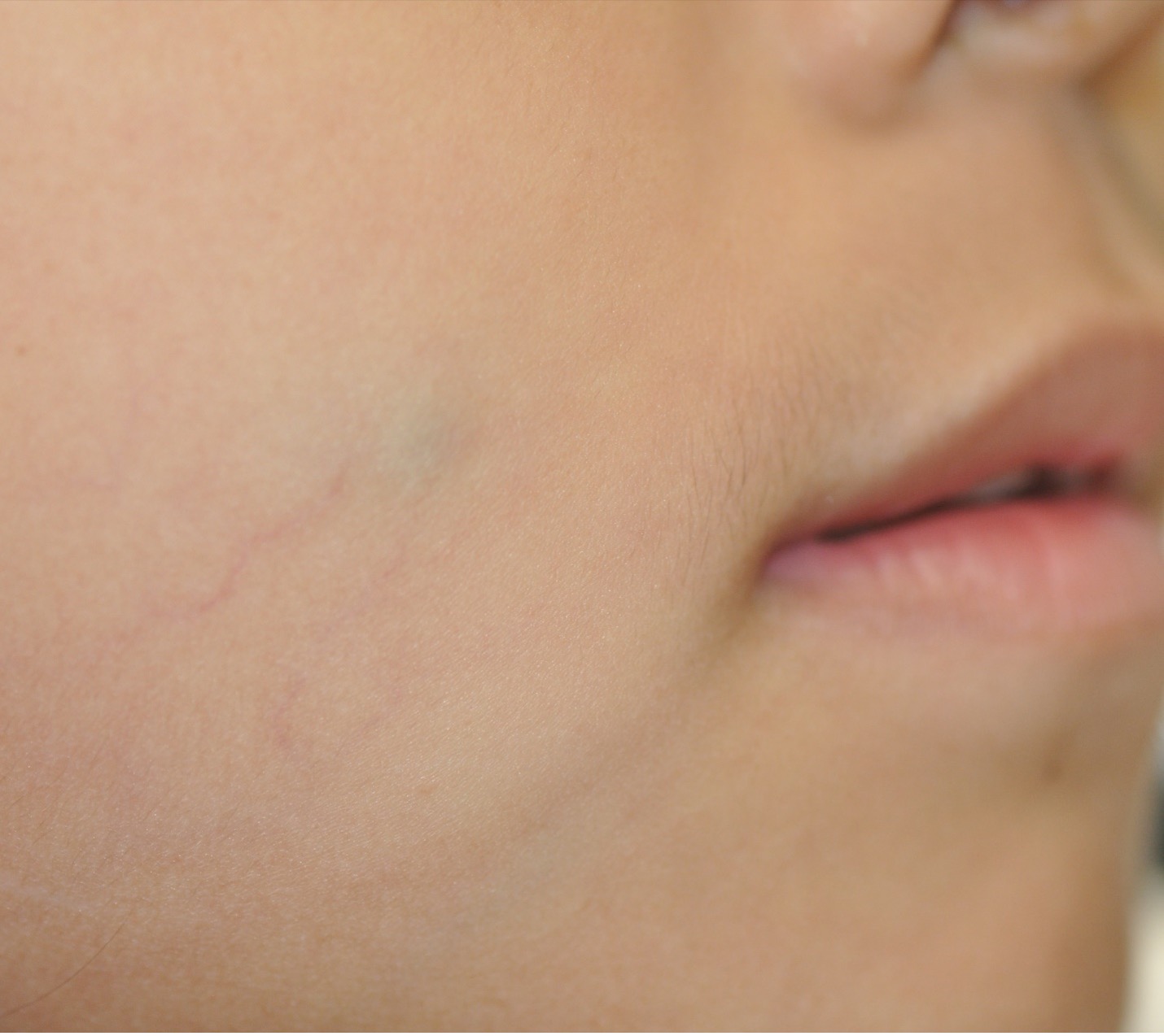
Figure 3.

Figure 4.

Figure 5.

Figure 6.
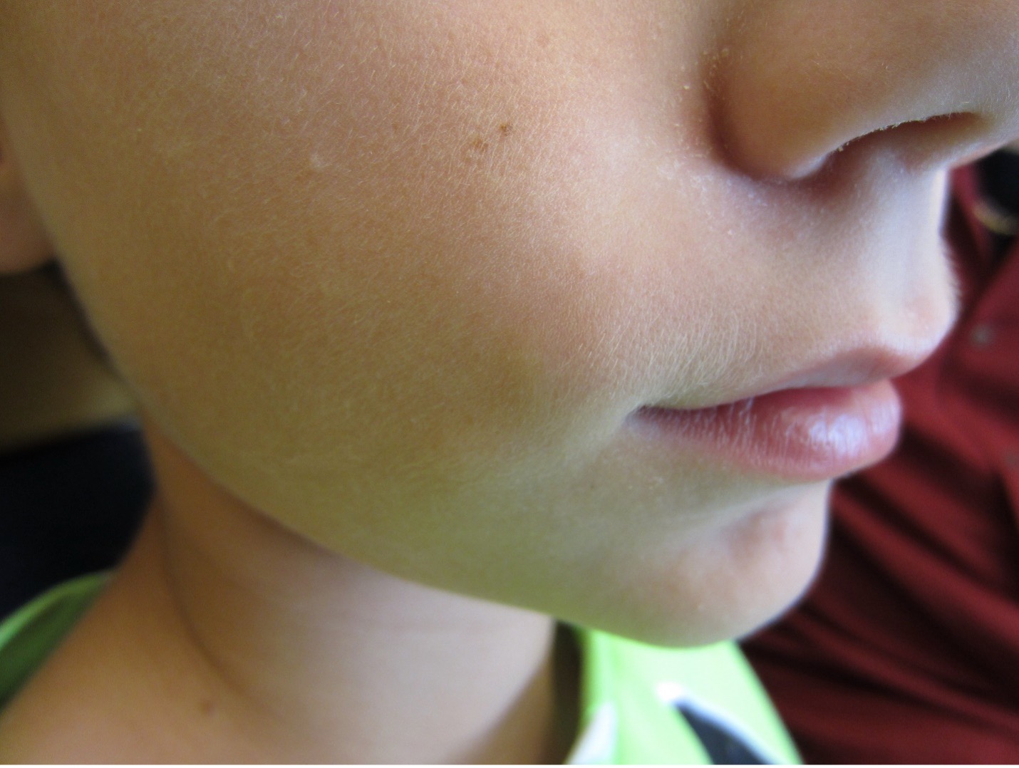

Figure 7.
Several clinical variants have been recognized. In the pseudobullous or anetodermic variant, the lesion is bullous-looking, and the overlying skin is atrophic, translucent, pink, or erythematous (Figure 8).7 Telangiectasis may be seen. The tumor is rapidly growing. Sites of predilection include the upper arms and shoulders.13 A pseudobullous or anetodermic pilomatricoma can be depressed at the center when vertical pressure is applied (dimple sign).13 Rarely, a pilomatricoma may rupture, resulting in an ulcerated or crusted nodule; this variant is referred to as perforating pilomatricoma (Figure 9).4,7 A pilomatricomal horn is a superficial variant of pilomatricoma.14 Giant pilomatricoma is another clinical variant, arbitrarily defined as a lesion greater than 5 cm.7

Figure 8.

Figure 9.
Most cases are sporadic. Multiple pilomatricomas occur in 2 to 5% of cases.1 The presence of 6 or more pilomatricomas is highly suggestive of an underlying disorder such as Gardner syndrome, Turner syndrome, Rubinstein-Taybi syndrome, Kabuki syndrome, Churg-Strauss syndrome, basal cell naevus syndrome (Gorlin syndrome), Soto syndrome, constitutional mismatch repair deficiency (CMMR-D), myotonic dystrophy, xeroderma pigmentosum, sarcoidosis, or trisomy.7,9,11,12,15-18 Although pilomatricoma is generally benign, malignant transformation has been, very rarely, described.19,20