
What’s Wrong With These Feet?

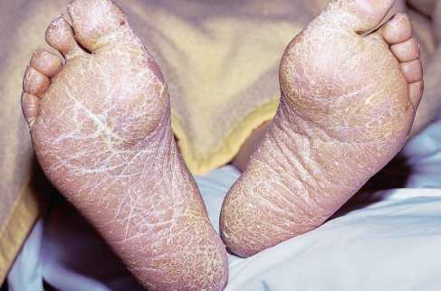
A 5-year-old boy was brought to his physician with a pruritic, erosive, and erythematous rash that appeared in a recurrent pattern on the bottom of his feet. Scaling and deep-seated (tapioca-like) vesicles were also observed.
What is the diagnostic picture here?
(Answer on next page.)
Answer: “Dyshidrotic” eczematous dermatitis
The characteristic lesions of “dyshidrotic” eczematous dermatitis can appear on soles, palms, and sides of toes and fingers. The disease origin is unknown; however, affected persons often have a family history of atopy—as this child did.
Treatment is symptomatic, aimed at controlling inflammation and relieving itching. It consists of high-potency topical corticosteroid therapy combined with a gel containing coal tar.
(Case and photograph courtesy of Dr Reynold C. Wong.)

A 28-year-old Mexican-American farm worker sought consultation for swelling, slight tenderness, and many small, draining sinus tracts on the bottom of her right foot; the exudate was bloody and purulent. The condition had been present for several months.
To what do you attribute the woman’s disorder?
(Answer on next page.)
Answer: Mycetoma
A clinical diagnosis of mycetoma was made; this was further substantiated when characteristic granular material was seen draining from the sinus tracts. Microscopic examination
revealed colonies of fungal hyphae, but cultures were negative except for one that showed aRhizopus species that had not previously been associated with mycetoma.
Mycetoma is a chronic infectious disease of the cutaneous and subcutaneous tissues, fascia, and bone; it generally affects the foot, although the hand and other sites may also be involved. It is caused by various species of fungi or Actinomyces. Mycetoma is relatively rare in the United States; it is more common in India, the Sudan, southern Asia, and the tropical regions of Central and South America, especially in rural areas where people go barefoot.
After 3 months of antibiotic therapy, the patient in this case seemed to be slowly improving, but the granules were still present in the draining sinus tracts. An x-ray study of the foot revealed no evidence of osteomyelitis. After 2 years, the patient’s condition had improved, but she was still experiencing chronic flare-ups.
(Case and photograph courtesy of Dr Reynold C. Wong.)
A 75-year-old man was admitted to the hospital because of weight loss (7 kg over the past 5 months) and easy fatigability. Physical examination revealed cachexia and hyperkeratosis of the soles of both feet. The patient said that the latter condition had developed within the past 5 months. Laboratory tests showed iron deficiency anemia, an elevated erythrocyte sedimentation rate (110 mm/h), and an elevated serum level of lactate dehydrogenase. An abdominal ultrasonogram revealed multiple small lesions on the liver, which were compatible with metastases.
Of what condition might the hyperkeratosis be a sign?
(Answer on next page.)
Answer: Adenocarcinoma
Gastroscopic examination showed a huge mass in the minor curvature; histologic examination proved it to be an adenocarcinoma. This is a rare case of adenocarcinoma of the stomach that first appeared clinically with a paraneoplastic manifestation: hyperkeratosis of the soles.
(Case and photograph courtesy of Drs N. K. Akritidis and K. G. Kistis.)
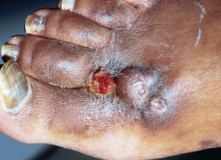
During the past 2 months, painless nodules had developed on a 48-year-old man’s right foot. His HIV status was initially unknown, but he reported being bisexual. Examination of his lower right leg revealed no erythema, edema, malodor, drainage, or warmth, and there were no palpable inguinal lymph nodes on that side. A preliminary diagnosis of pyogenic granuloma was made.
What other condition could have caused the nodules?
(Answer on next page.)
Answer: Kaposi sarcoma
A 4-mm punch biopsy revealed the lesions to be the nodular form of Kaposi sarcoma. Suspicion that
the patient was HIV-positive was confirmed by Western blot test and enzyme-linked immunosorbent assay.
Skin biopsy is imperative to differentiate Kaposi sarcoma from the strikingly similar bacillary angiomatosis, an infectious disease that is also common among patients with AIDS.
(Case and photograph courtesy of Drs Bryan Caldwell and Donald Kushner.)