
Hutchinson-Gilford Progeria Syndrome
Authors:
Danielle R. Bullock, MD
Division of Pediatric Rheumatology, Department of Pediatrics, University of Minnesota Medical School, Minneapolis, Minnesota
Joseph Sameji, MD, and Elena Mantz, MD
Selian Lutheran Hospital, Arusha, Tanzania
Emily Hall, DO
Providence St. Joseph Medical Center, Polson, Montana
Citation:
Bullock DR, Sameji J, Mantz E, Hall E. Hutchinson-Gilford progeria syndrome [published online November 8, 2017]. Consultant360.
At a clinic in the East African country of Tanzania, a 4-year-old boy presented for evaluation of scrotal swelling. The boy appeared underweight and stunted. He had been of normal weight until 4 months of age, followed by decelerated weight velocity with an abrupt plateau. When plotted on growth charts, his height and weight were less than the first percentile, and his occipital-frontal circumference (OFC) was at the first percentile (Figures 1a, 1b, and 1c).


Figure 1. Growth charts showed that the patient's height and weight were less than the first percentile, and his OFC was at the first percentile.
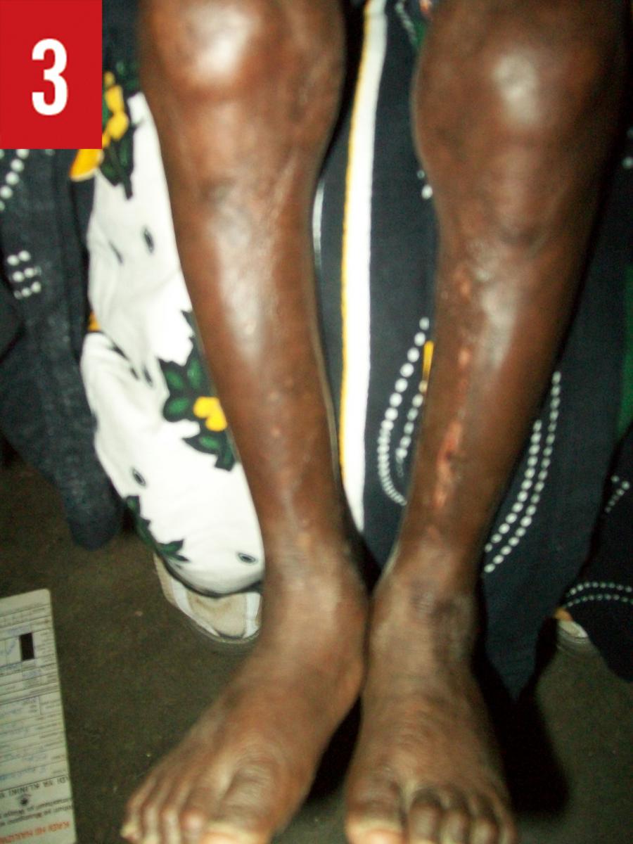
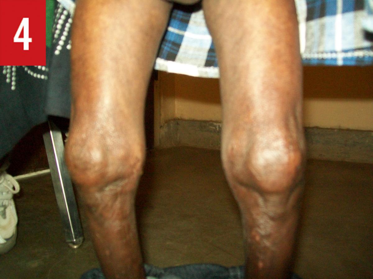
Physical examination. His head was large, with frontal bossing and an open anterior fontanelle. In a high-pitched voice, he answered questions appropriately for his age and could count to 10 in several languages. He had minimal subcutaneous fat, thin and brittle nails, hypotrichosis with prominent scalp veins, absent eyebrows with minimal eyelashes, absent ear lobes but well-formed pinnae, crowded teeth, and a protuberant, soft abdomen. Inspection of the skin revealed hyperpigmented, nonblanchable patches on the trunk (Figure 2) and taut skin over the shins (Figure 3). He had a stiff handgrip, contractures of the fifth fingers, and deformity of the knees with limitation in flexion and extension (Figure 4). Movement of the elbows, wrists, ankles, and spine were limited, while his hips, shoulders, and neck had normal range of motion. His gait was wide-based. Strength and tone were normal. Genitourinary examination findings were notable for a nontender mass that transilluminated, suggestive of a hydrocele. His vital signs were normal.

History. His medical history was notable for premature birth at 35 weeks, at which time he had weighed 2 kg, which was appropriate for gestational age. He had been hospitalized for 2 weeks following his birth due to poor feeding. Since then, he had not been hospitalized. He had been exclusively breastfed until age 6 months, at which time solid foods had been introduced. Supplemental breastfeeding had continued until age 2 years. Tooth eruption had occurred at age 3 years.
He had normal gross motor, fine motor, and verbal development. There was no family history of poor weight gain or short stature and no history to suggest food insecurity. Both parents identified ethnically as Maasai. There was no consanguinity.
Diagnostic tests. Due to limited resources, the diagnostic workup was limited. The patient demonstrated growth failure of weight and height with relative sparing of OFC, a finding consistent with type II failure to thrive. His family growth history excluded familial short stature or constitutional growth delay. Hypothyroidism seemed unlikely, given his cognitive function, but an endocrine abnormality remained possible. Ultrasonography of the head confirmed an open anterior fontanelle but no other abnormalities.
The constellation of his clinical features—growth failure, relative sparing of head size, normal intelligence, and musculoskeletal and dermatologic abnormalities—raised suspicion for Hutchinson-Gilford progeria syndrome (HGPS). Results of genetic testing were positive for a heterozygous mutation in exon 11 of the lamin A/C gene (LMNA), confirming the diagnosis.
Discussion. HGPS is a rare genetic syndrome affecting 1 in 4 million live births.1 Children with this condition develop atherosclerosis, kidney failure, loss of eyesight, and cardiovascular problems, and they face atherosclerotic-related death at a mean age of 14 years.1-3
There is no cure for HGPS. Treatment focuses on cardiovascular health, specifically the slowing of atherosclerotic disease.3
Outcome of the case. The patient is now 7 years old. He has been without any recognized ischemic or cardiac events, but he does have progressive arthritis that affects his daily functioning.
References:
- Pollex RL, Hegele RA. Hutchinson-Gilford progeria syndrome. Clin Genet. 2004;66(5):375-381.
- LMNA gene. Genetics Home Reference. https://ghr.nlm.nih.gov/gene/LMNA. Accessed November 7, 2017.
- About progeria. Progeria Research Foundation. http://progeriaresearch.org/about_progeria. Accessed November 7, 2017.
Funding/Support:
The Progeria Research Foundation paid for the patient’s genetic testing. No funding was secured for the writing of this case report.