
Interactive Quiz: Forceful Vomiting in a Newborn

Welcome to Gastroenterology Consultant's latest interactive diagnostic quiz. Over the next few pages, we'll present a case and ask you to make the diagnosis and treat the patient. Along the way, we'll provide details about the case, and at the end, we'll share the patient's outcome.
First, let’s meet the patient.
A 3-week-old neonate, born at 37 3/7 weeks’ gestational age, presented to a clinic after 5 days of projectile, nonbilious emesis and poor weight gain. His toddler sibling had just recovered from an acute vomiting illness after foreign travel.
The neonate’s physical examination findings at the time were normal, and no masses were palpable in the abdomen. An abdominal ultrasonogram showed a pylorus with normal measurements and gastric contents passing through in real-time imaging. Electrolyte levels were normal.
One week later, persistent vomiting had led to a 6% loss of body weight, and the patient was admitted to the hospital.
Answer: a. Perform a second abdominal ultrasonogram
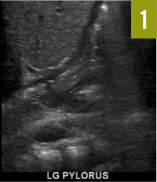
A second abdominal ultrasonogram, performed 1 week after the initial examination in our clinic, showed a pylorus with normal wall thickness (3.0 ´ 14.4 ´ 9.8 mm, Figure 1). The pylorus was opening widely intermittently, and gastric contents were passing through. A kidney, ureter, and bladder radiograph demonstrated a normal gas pattern. An upper gastrointestinal radiographic series showed a markedly dilated stomach, reflux of stomach contents into the esophagus, and delay in gastric emptying, but no anatomic abnormalities (Figure 2).

Answer: b. Perform upper GI endoscopy
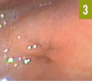
The patient then underwent upper gastrointestinal endoscopy, which demonstrated a narrow opening to the pylorus, edematous pyloric folds with mucosal hypertrophy, and antral nippling (protrusion into the stomach; Figure).
How would you diagnose this condition?
Answer: b. Infantile hypertrophic pyloric stenosis
Infantile hypertrophic pyloric stenosis (IHPS) is a common cause of gastrointestinal tract obstruction, occurring in approximately 2 to 3 neonates per 1000 births.2 Classically, IHPS presents between 4 and 8 weeks of life3 with forceful nonbilious, nonbloody emesis, a palpable olive-like mass in the abdomen, hypochloremia with metabolic alkalosis, and ultrasonography findings of a thickened pylorus. However, in this case, no olive-like mass was palpable, and the patient had 2 abdominal ultrasonograms that showed normal results.
This case illustrates that IHPS should remain in the differential diagnosis for newborns with forceful vomiting despite normal abdominal examination findings and normal ultrasonography findings.
How would you treat this patient?
Answer: The patient received definitive surgical treatment by pyloromyotomy.
Our patient’s hypochloremia was an important clinical clue. Electrolyte abnormalities are only observed in approximately half of infants with IHPS. One study that compared infants with IHPS with infants with gastroesophageal reflux disease determined that the sensitivities of hypochloremia (≤98 mEq/L) and metabolic alkalosis (≥29 mEq/L) were low (approximately 50% and 36%, respectively). However, their presence was highly predictive (97% and 96%, respectively) and highly specific (both 99%) for IHPS.8 Therefore, when clinical symptoms strongly suggest IHPS, we advocate for the use of clinical judgment for prompt referral for surgical management. If confirmation of IHPS is desired in inconclusive cases to avoid unnecessary surgery, upper gastrointestinal endoscopy allows direct visualization of the hypertrophied pylorus.9
Outcome of the case:
Subsequently, the patient tolerated oral feedings and was discharged home.
Authors:
Shiela Beroukhim
David Geffen School of Medicine at UCLA, Los Angeles, California
George Gershman, MD
Department of Pediatrics, Division of Gastroenterology, Harbor–UCLA Medical Center, Torrance, California
Jennifer K. Yee, MD
Department of Pediatrics, Division of Pediatric Endocrinology, Harbor–UCLA Medical Center, Torrance, California
Citation:
Beroukhim S, Gershman G, Yee JK. Infantile hypertrophic pyloric stenosis [published online February 27, 2018]. Consultant for Pediatricians. https://www.consultant360.com/exclusives/infantile-hypertrophic-pyloric-stenosis.
References:
- Costa Dias S, Swinson S, Torrão H, et al. Hypertrophic pyloric stenosis: tips and tricks for ultrasound diagnosis. Insights Imaging. 2012;3(3):247-250.
- Krogh C, Fischer TK, Skotte L, et al. Familial aggregation and heritability of pyloric stenosis. JAMA. 2010;303(23):2393-2399.
- Aboagye J, Goldstein SD, Salazar JH, et al. Age at presentation of common pediatric surgical conditions: reexamining dogma. J Pediatr Surg. 2014;49(6):995-999.
- van der Schouw YT, van der Velden MT, Hitge-Boetes C, Verbeek AL, Ruijs SH. Diagnosis of hypertrophic pyloric stenosis: value of sonography when used in conjunction with clinical findings and laboratory data. AJR Am J Roentgenol. 1994;163(4):905-909.
- Godbole P, Sprigg A, Dickson JA, Lin PC. Ultrasound compared with clinical examination in infantile hypertrophic pyloric stenosis. Arch Dis Child. 1996;75(4):335-337.
- Macdessi J, Oates RK. Clinical diagnosis of pyloric stenosis: a declining art. BMJ. 1993;306(6877):553-555.
- Keckler SJ, Ostlie DJ, Holcomb GW III, St Peter SD. The progressive development of pyloric stenosis: a role for repeat ultrasound. Eur J Pediatr Surg. 2008;18(3):168-170.
- Smith GA, Mihalov L, Shields BJ. Diagnostic aids in the differentiation of pyloric stenosis from severe gastroesophageal reflux during early infancy: the utility of serum bicarbonate and serum chloride. Am J Emerg Med. 1999;17(1):28-31.
- Liacouras CA, Cook-Sather SD, Schreiner MS, Bellah RD. Endoscopic findings in hypertrophic pyloric stenosis: appearance in classic and evolving disease. Gastrointest Endosc. 1997;45(5):371-374.