
Osteochondral Fracture in a Baseball Player
A 12-year-old boy presented with pain in the right shoulder. Six months earlier, he had fallen on his outstretched right hand during baseball practice and had persistent shoulder pain ever since.
 The patient had full range of motion, but he complained of pain with abduction of his right arm. Tenderness was noted over the anterior portion of the shoulder joint capsule. There was also generalized muscle weakness in the right arm but no evidence of decreased muscle bulk. The weakness was attributed to decreased effort by the patient as a result of the pain.
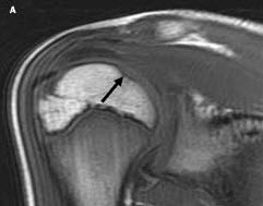
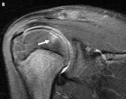
MRI of the right shoulder showed a subchondral crescent-shaped area of signal abnormality on the superomedial portion of the humeral head (A, black arrow) along with subchondral edema (B, white arrow). The appearance of the signal abnormality, along with the underlying edema, is consistent with an osteochondral fracture of the humeral head.
 Osteochondral fractures, also known as osteochondritis dissecans, frequently occur in children and have no gender predilection. Symptoms include the inability to bear weight and intense pain, which typically decreases within 2 or 3 weeks of the original injury. Osteochondral fractures are primarily caused by either ischemia or trauma, which may be direct or repetitive microtrauma. They are most common in the weight-bearing areas and are often seen in a femoral condyle and the talus. When the fracture occurs in the shoulder joint, it usually involves either the humeral head or the glenoid.
Treatment typically consists of splint application and subsequent immobilization of the joint. Resting of the injured extremity along with NSAIDs can reduce pain severity. Surgery is occasionally necessary to remove the intra-articular loose body.
(Case and photographs courtesy of Douglas Beall, MD, of Edmond, Okla, and John Whyte, MD, of Silver Spring, Md.)