
Calcified Splenic Cyst
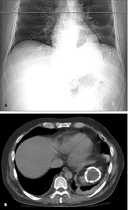
A 38-year-old man found lying on the floor in his home was hospitalized because of alcohol intoxication. A chest radiograph showed a large calcified lesion in the left upper abdomen (A). A CT scan with intravenous contrast revealed a large, well-defined, cystic mass with mural calcification in the spleen (B). The CT findings were not consistent with a vascular malformation or echinococcal cyst—specifically, the mass was sharply demarcated, unilocular without septations, and round with a thin wall and attenuation similar to water. Urine Histoplasma antigen test results were negative.
The patient underwent CT-guided aspiration of the cyst, and the fluid was cultured and examined under microscopy. Culture, cytology, and Gram stain results were negative, and no parasitic elements were identified under microscopy. Based on these results and the lack of a patient history of pancreatitis, traumatic calcified splenic cyst was diagnosed.
Calcified splenic cysts most often occur in patients who are prone to recurrent blunt abdominal trauma, such as alcoholics. However, this is a diagnosis of exclusion; more common, treatable causes of splenic masses need to be ruled out first.