
What is Causing this Woman's Dermal Nodules and Clubbing?
AUTHORS:
Mackenzie Gwynne, AB
University of South Carolina School of Medicine, Columbia, South Carolina
Barbara B. Wilson, MD
Department of Dermatology, University of Virginia, Charlottesville, Virginia
A 58-year-old woman with Graves disease complained of a 4-year history of asymptomatic nodules on her toes and thickening of her shins. The large toes’ nodules were painful when she wore shoes, necessitating her wearing men’s shoes.
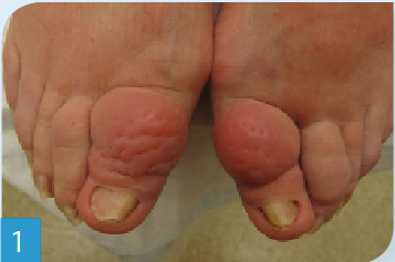
Upon physical examination, the patient had firm erythematous symmetric dermal nodules involving the dorsal aspects of both great toes (Figure 1), as well as erythema and thickening of her shins extending circumferentially onto her calves (Figures 2 and 3). The patient also had clubbing of her fingers. There was no presence of ophthalmopathy.

Answer: Pretibial myxedema and thyroid acropachy
A clinical diagnosis of pretibial myxedema and thyroid acropachy was made; however, ophthalmopathy was not present. She was instructed to apply clobetasol ointment to her shins daily, using Saran Wrap occlusion for the first 10 days. One half cubic centimeter of Kenalog 40 mg/cc was injected into each toe nodule. Two months later, she had remarkably reduced size and softening of the nodules on her toes as well as of her shins (Figure 4). She reported that she no longer had to wear men’s shoes. Repeat Kenalog injections were performed and she was to continue the clobetasol without Saran Wrap occlusion until adequate improvement was attained.

Discussion. Pretibial myxedema is a rare manifestation of Graves disease and can also occur in patients with Hashimoto’s thyroiditis.4 Pretibial myxedema manifests clinically as bilateral nonpitting thickening and induration of the skin with well-demarcated dermal nodules.4 The lesions may have a violaceous, erythematous, or a yellow-brown pigment with a peau d’orange appearance. The anterolateral aspects of the shins are normally involved, but more extensive involvement of the lower legs may occur. Involvement of the dorsa of the feet, elbows, knees, and neck have been reported less commonly.3 Lesions are usually asymptomatic but pruritus and pain have been described by some patients. The diagnosis of pretibial myxedema is based on history and clinical appearance; however, punch biopsy may be necessary to confirm the diagnosis in some cases.5 The finding of pretibial myxedema is associated with a high percentage of patients who also have ophthalmopathy.1 Ophthalmopathy usually presents first and is followed in severe cases by thyroid dermopathy such as pretibial myxedema and/or thyroid acropachy.5 Pretibial myxedema is due to glycosaminoglycans (GAG) in the dermis specifically hyaluronic acid.3 Thyroid stimulating hormone (TSH) receptor antibodies are thought to be responsible for recognizing connective tissue antigens in the skin and behind the eyes producing thyroid dermopathy and ophthalmopathy, respectively.1 Both humoral and cellular immune mechanisms are involved in triggering fibroblasts to produce a large amount of GAG.5 The pretibial area is usually involved due to mechanical factors and dependent position.5 Thyroid acropachy presents with digital clubbing, swelling, and periosteal reaction of the phalanges.6 It is almost always associated with ophthalmopathy, thyroid dermopathy, or both and is an indicator for the severity of the underlying autoimmune process.1
Treatment. Treatment is indicated when lesions are symptomatic or when there is a cosmetic concern. Mild cases of pretibial myxedema do not require treatment. Complete remission occurs in mild forms of the disease in 50% of the patients after several years.5 For more severe cases, the mainstay of treatment is medium-to-high potency topical corticosteroids with or without occlusive dressing.5 Intralesional corticosteroids may be used for patients who do not show improvement with topical agents.
References:
1. Ai J, Leonhardt JM, Heymann WR. Autoimmune thyroid diseases: etiology, pathogenesis, and dermatologic manifestations. J Am Acad Dermatol. 2003;48(5):641-659.
2. Doshi DN, Blyumin ML, Kimball AB. Cutaneous manifestations of thyroid disease. Clin Dermatol. 2008;26(3):283-287.
3. Fatourechi V. Pretibial myxedema: pathophysiology and treatment options. Am J Clin Dermatol. 2005;6(5):295-309.
4. Fatourechi V. Thyroid dermopathy and acropachy. Best Pract Res Clin Endocrinol Metab. 2012;26(4):553-565.
5. Fatourechi V, Ahmed DD, Schwartz KM. Thyroid acropachy: report of 40 patients treated at a single institution in a 26-year period. J Clin Endocrinol Metab. 2002;87(12):5435-5441.