
What is the Cause of this Man's Erosive Nodule?
A 77-year-old white man presented for evaluation of an erythematous and erosive nodule of the umbilicus (Figure 1). He complained of abdominal pain and nausea. A biopsy was performed (Figures 2 and 3). His medical history is significant for coronary artery disease, treated by bypass grafting, prostate cancer treated by radium seed implants, and bowel obstruction due to carcinoma of the small intestine and omentum treated 1 year previously. His mother had a history of colon cancer. Pathology included both a carcinoembryonic antigen and CDX2 stain. The patient was scheduled for a carcinoembryonic antigen (CEA) and Cdx2 stain, as well as a CT scan and biopsy.

What's Your Diagnosis?
(Answer and discussion on next page)
Answer: Sister Mary Joseph’s Nodule
The biopsy specimen revealed strands of neoplastic cells extending between thickened collagen bundles. Nuclear pleomorphism is noted and individual cell necrosis is identified. A CEA stain is strongly positive and decorates neoplastic cells extending to the margins of the biopsy specimen (Figure 4). A CDX-2 stain also decorates neoplastic cells (Figure 5). The histologic findings are those of carcinoma cutis, and support the clinical impression of metastatic adenocarcinoma of gastrointestinal origin in the form of a Sister Mary Joseph’s nodule (SMJN).
Discussion
SMJN is an umbilical metastasis associated with intra-abdominal and intrapelvic disease.1 Named for Sister Mary Joseph, the first to correlate the link between the eponymous umbilical nodules and intra-abdominal disease, the nodule presents as a firm indurated lesion, whose surface may be fissured, ulcerated, or necrotic.2 Comparable to other cutaneous metastases, the SMJN itself is often painless, yet contributes to internal symptoms (ie, nausea, vomiting, dyspepsia, abdominal pain, or weight loss).2
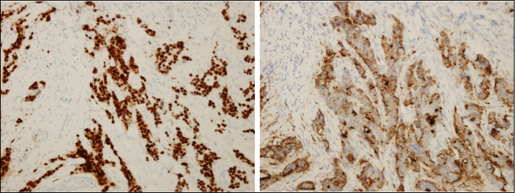
Cutaneous metastases, like the SMJN, can often present histological patterns characteristic of the original, underlying tumor. In such cases, diagnosis must center on histopathologic evaluation, in which the use of immunohistochemical studies can be very helpful. In this specific case, a carcinoembryonic antigen (CEA) and CdX2 stains were invaluable in tracing the origin of the SMJN. The CEA stain was strongly positive, which pointed toward a colorectal origin.3 The further use of a CdX2 stain, a recently cloned homeobox gene which serves as an even more specific and highly sensitive marker for metastases of a gastrointestinal origin, was also positive, and decorated the neoplastic cells.4 This staining panel greatly aided in the final diagnosis’ proposed origin.
The other entities in the differential diagnosis can be excluded by physical examination and biopsy with histopathological review.
Outcome of the Case
The CT scan of our patient revealed extensive intra-abdominal and hepatic metastases. An oncology consult recommended a course of chemotherapy for the patient, however treatment was never initiated. Tumor progression and intra-abdominal and hepatic metastases will be monitored closely.
The author discusses their case in the podcast below:
REFERENCES:
- Fairchild A, Janoski M, Dundas G. Sister Mary Joseph’s nodule. CMAJ. 2007;176(7):929-930.
- Abu-Hilal M, Newman JS. Sister Mary Joseph and her nodule: historical and clinical perspective. Am J Med Sci. 2009;337(4):271-273.
- Wong CY, Helm MA, Kalb RE, et al. The presentation, pathology, and current management strategies of cutaneous metastasis. N Am J Med Sci. 2013;5(9):499-504.
- Bayrak R, Haltas H, Yenidunya S. The value of CDX2 and cytokeratins 7 and 20 expression in differentiating colorectal adenocarcinomas from extraintestinal gastrointestinal adenocarcinomas: cytokeratin 7-/20+ phenotype is more specific than CDX2 antibody. Diagn Pathol. 2012;7:9.
- Powell FC, Su WP, Perry HO. Pyoderma gangrenosum: classification and management. J Am Acad Dermatol. 1996;34(3):395-409.
- Skin—Nonmelanocytic tumors. Pathology Outlines’s Web site. PathologyOutlines.com. http://pathologyoutlines.com/topic/skintumornonmelanocyticmucinouscarcinoma.html. Updated June 2012. Accessed July 20, 2014.
- Elder DE. Pathology of Melanoma. Clin Cancer Res. 2006;12(7 Pt 2);2308s-2311s.
- Smoller BR. Histologic criteria for diagnosing primary cutaneous malignant melanoma. Mod Pathol. 2006;19 Suppl 2:S34-S40.