
Popsicle Panniculitis
 The parents of a 9-month-old boy were concerned about the enlarging areas of reddish discoloration on his cheeks. The infant was born at full term and had been healthy, with no significant medical or family history; specifically, he had no bug bites, trauma, illnesses, or sick contacts. His growth and development were appropriate and immunization status was up-to-date.
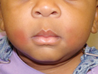
The afebrile, playful infant had symmetrical erythematous, indurated, nontender lesions of 2 × 2 cm on his cheeks close to the angle of the mouth. The overlying skin was intact, with no associated warmth, and the oral cavity was normal. All other physical findings were unremarkable.
Further questioning revealed that 2 days before the lesions appeared, the mother had given the infant a popsicle for teething. The classic history and presentation led to a clinical diagnosis of popsicle panniculitis—a benign, self-limited form of cold panniculitis.
Popsicles, icepacks, and exposure to cold have all been shown to cause cold panniculitis in children. It predominantly occurs during infancy after cold injury and generally affects the cheeks and chin—areas that are rich in subcutaneous fat and more often exposed to cold. Skin sensitivity to cold diminishes markedly with age.1 Lemez2 showed that ice applied to the skin for 50 seconds resulted in a subcutaneous nodule in 100% of newborns, in 40% of infants younger than 6 months, and in only a few infants younger than 9 months.
The underlying mechanism is thought to be the higher concentration of saturated fatty acids in the subcutaneous fat of neonates3 that crystallize after a slight decrease in body temperature.4 The subcutaneous fat in adults has predominantly unsaturated fats, which may explain why popsicle panniculitis occurs almost exclusively in children. Histopathologically, it is a lobular panniculitis5 characterized by intense inflammation at the dermosubcutaneous junction.
Popsicle panniculitis usually presents as areas of reddish discoloration or as red-purple, indurated,
nontender swellings of the cheeks or submental region 24 to 48 hours after contact with a popsicle or ice cube. The diagnosis is clinical. Facial cellulitis, trauma, and frostbite usually
can be differentiated from cold panniculitis by means of a careful history and physical examination.
The outcome is excellent, with spontaneous and complete resolution over several weeks. The lesions may recur and can be prevented by avoiding cold exposure and direct contact with ice products. Caregivers can be educated and reassured regarding the benign nature of the condition.
1. Duncan WC, Freeman RG, Heaton CL. Cold panniculitis. Arch Dermatol. 1966;94:722-724.
2. Lemez L. Beitrag zur pathogenese der subcutanen fettgewebsnekrose neugeborener (sog. Sclerodermia neonatorum) an der hand einer kaltereaktion des subcutanen fettgewebes bei neugeborenen und jungen sauglingen. Z Kinderheilk. 1928;46:323-369.
3. Hirsch J. Fatty acid patterns in human adipose tissue. In: Handbook of Physiology. Baltimore: Williams and Wilkins; 1965:181-190.
4. Collins HA, Stahlman M, Scott HW Jr. The occurrence of subcutaneous fat necrosis in an infant following induced hypothermia used as an adjuvant in
cardiac surgery. Ann Surg. 1953;138:880-885.
5. Requena L, Sánchez Yus E. Panniculitis. Part II. Mostly lobular panniculitis. J Am Acad Dermatol. 2001;45:325-361.