
A Lupus-like Eruption During Gemcitabine Therapy for Metastatic Breast Carcinoma
A 69-year-old Caucasian woman with metastatic breast carcinoma demonstrated good clinical response to gemcitabine therapy with reduction in her metastatic load. She developed a widespread papulosquamous eruption shortly after starting gemcitabine therapy. Her skin biopsy and clinical findings supported a diagnosis of cutaneous lupus erythematosus. Because of the relatively minor symptoms from her skin disease, she was able to remain on therapy.
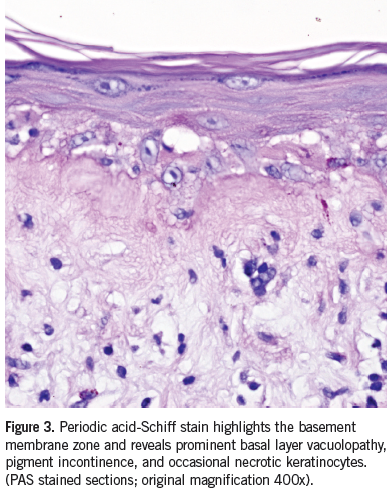
Clinical course. At the start of her treatment regimen, our patient immediately developed intense erythematous annular scaling plaques of the bilateral arms, chest, abdomen, back, and thighs with exfoliation of the soles of the bilateral feet (Figure 1). She was given clobetasol cream with no relief. An antinuclear antibody (ANA) revealed a titer of 1:640 in a speckled pattern. Biopsy revealed an atrophic epidermis associated with basal layer vacuolopathy consistent with cutaneous lupus erythematosus (Figures 2 and 3). Direct immunoflourescence also revealed changes consistent with cutaneous lupus erythematosus.1 She began taking diphenhydramine prior to her gemcitabine therapy and was also placed on a prednisone taper at the time of her gemcitabine treatment. The erythematous scaly patches and plaques completely resolved within a few days of the prednisone therapy. She subsequently completed her gemcitabine chemotherapy and had a significant decrease in her metastatic load 5 months after the start of treatment.

Overview of drug-induced lupus. Drug-induced lupus has been reported as a side-effect of long-term therapy with many medications.2 The most common agents are procainamide and hydralazine.3 Drugs less frequently associated with the disease are chlorpromazine, isoniazid, methyldopa, penicillamine, quinidine, and sulfasalazine.3 Gemcitabine has recently been associated with subacute lupus erythematosus.4
Lupus, itself, is a chronic inflammatory autoimmune disease divided into 4 main types: systemic lupus erythematousus (SLE), neonatal, discoid, and drug-induced.5 Drug-induced lupus erythematosus refers to a condition whose clinical, histological, and immunological features are similar to those seen in idiopathic lupus erythematosus.6 There is no set criteria for drug-induced lupus, however 4 most common features including arthritis, serositis, ANA, and antihistone antibodies are present after the initiation of treatment with a drug and must resolve after discontinuation.7
Cutaneous Side Effects. Gemcitabine is a nucleoside analog used as a chemotherapeutic agent. It is generally well-tolerated with rare side effects including myelosuppression, GI disturbances, influenza-like symptoms, elevated liver transaminases enzymes, peripheral edema, and mild monomorphic exanthematous rash.8 Specific to the skin, gemcitabine has been reported to cause a phototoxic recall reaction which each subsequent dose of gemcitabine.9 Scleroderma-like changes including inflammatory edema in the lower extremities and biopsy revealing diffuse sclerosis without involvement of fascia or muscle, tightening, and induration of the skin on the trunk and extremities has also been reported.10 Acro-necrosis of the upper limbs after gemcitabine administration has also been reported.11
Management Strategies. Drug-induced lupus typically resolves after discontinuation of the offending agent. In many instances, such as for chemotherapeutic agents, stopping the drug is not an option. Previous reports combining gemcitabine with a short course of high-dose prednisone, 60 mg/day tapered over 3 days plus topical triamcinolone 0.1% ointment allowed a safe re-challenge with subsequent doses of gemcitabine.8 We recommend a prednisone taper beginning at 30 mg daily for 10 days, 20 mg daily for 10 days, and followed by 10 mg daily for 10 days.
Conclusion. There are an estimated 15,000 to 30,000 cases of drug-induced lupus per year.12 The most common cutaneous manifestation is a butterfly rash across the bridge of the nose and cheeks while sparing the nasaolabial folds. However, atypical manifestations such as erythematous annular exfoliative plaques may be seen. If drug-induced lupus erythematosus is suspected, we recommend incisional biopsy with hematoxylin and eosin staining, along with direct immunoflourescence for deposits of IgG to confirm the diagnosis. When discontinuation of a drug is not an option, we recommend a prednisone taper.
References:
1. Ramdial PK, Naidoo DK. Drug-induced cutaneous pathology. J Clin Pathol. 2009;62(6):493-504.
2. Rubin RL. Drug-induced lupus. Toxicology. 2005;209(2):135-147.
3. Price EJ, Venables PJ. Drug-induced lupus. Drug Saf. 1995;12(4):283-290.
4. Wiznia LE, Subtil A, Choi JN. Subacute cutaneous lupus erythematosus induced by chemotherapy: gemcitabine as a causative agent. JAMA Dermatol. 2013;149(9):1071-1075.
5. Maidhof W, Hilas O. Lupus: an overview of the disease and management options. P T. 2012;37(4):240-249.
6. Pretel M, Marques L, Espana A. Drug-induced lupus erythematosus. Actas Dermosifiliogr. 2014;105(1):18-30.
7. Antonov D, Kazandjieva J, Etugov D, et al. Drug-induced lupus erythematosus. Clin Dermatol. 2004;22(2):157-166.
8. Katsenos S, Nikolopoulou M. Gemcitabine-induced severe peripheral edema in a patient with lung cancer. J Pharm Pract. 2012;25(3):393-395.
9. Badger J, Kang S, Uzieblo A, Srinivas S. Double diagnosis in cancer patients and cutaneous reaction related to gemcitabine: CASE 3. Photo therapy recall with gemcitabine following ultraviolet B treatment. J Clin Oncol. 2005;23(28):7224-7225.
10. Bessis D, Guillot B, Legouffe E, Guilhou JJ. Gemcitabine-associated scleroderma-like changes of the lower extremities. J Am Acad Dermatol. 2004;51(Suppl):S73-S76.
11. D'Alessandro V, Errico M, Varriale A, et al. [Case report: Acro-necrosis of the upper limbs caused by gemcitabine therapy]. Clin Ter. 2003;154(3):207-210.
12. Borchers AT, Keen CL, Gershwin ME. Drug-induced lupus. Ann NY Acad Sci. 2007;1108:166-182.