
Interactive Quiz: Slowly Growing Nodular Lesion

Welcome to Consultant360's new interactive diagnostic quiz. Over the next few pages, we'll present a case and ask you to make the diagnosis. Along the way, we'll provide details about the case, and at the end, we'll share the patient's outcome.
First lets meet the patient…

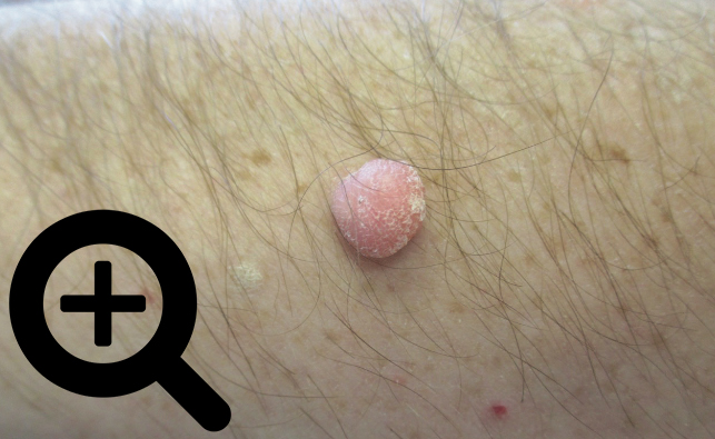
A 47-year-old man presented with an asymptomatic, slow-growing, nodular lesion on the left arm. He had first noted it approximately 7 years ago as a pea-sized lesion, and it had gradually increased to the present size. There was no history of ulceration or discharge from the lesion or trauma or insect bite in the area. The patient’s medical history was unremarkable. No other family members were affected with similar lesions.
Physical examination revealed a soft, skin-colored, nontender, well-defined, discrete, dome-shaped nodule on the lateral aspect of the left arm. No other skin abnormalities were noted. In particular, there were no comedo-like lesions, café au lait spots, or excessive hair growth on the surface of the lesion. There was no axillary lymphadenopathy. The rest of the physical examination findings were unremarkable.

Answer: Nevus Lipomatosus Superficialis (NLS)
Excisional biopsy of the lesion was performed and revealed globules of mature adipocytes around blood vessels and eccrine glands in the papillary and reticular dermis, along with hyperkeratosis and acanthosis of the overlying epidermis, confirming the clinical diagnosis of nevus lipomatosus superficialis.
Answer: NLS is a hamartoma that is characterized by the presence of ectopic mature adipose tissue in the dermis.1
Hoffmann and Zurhelle in 1921 were the first to report this skin anomaly.2 They described the case of a 25-year-old man with multiple congenital nodules in the left gluteal region and termed this entity naevus lipomatodes cutaneus superficialis. The condition is classified into 2 clinical types: the classical multiple type (also known as the Hoffmann-Zurhelle type) and the solitary type.3,4
Answer: NLS is an uncommon condition.
The exact incidence is not known, with the literature on this condition limited to case reports and case series. Suffice it to say, nevus lipomatosus superficialis is an uncommon condition. In a retrospective study, 8 cases were seen in an 11-year-period from 2001 to 2011 at a facility in Chandigarh, India.5 In another retrospective study, 8 cases were seen in a 14-year-period from January 1997 to December 2010 at a hospital in Tunis, Tunisia.6 The classical type is usually present at birth or may emerge in the first 2 decades of life.4,7 The solitary type, on the other hand, often appears later in life between the third and sixth decades.8,9There is no familial or gender predilection.4,7,10,11
Answer: No intervention is necessary, but surgery and injections are treatment options.
The condition is benign, without systemic abnormalities or malignant potential.4,7,10 Postsurgical recurrence, although rare, has been reported.6,22
Treatment is usually not necessary apart from reassurance and watchful observation, unless the lesion is symptomatic, the diagnosis is in doubt, or there is a cosmetic concern.7,12 Complete surgical excision is the treatment of choice for the solitary lesion.6,11,12 Surgical excision allows histologic examination of the lesion. However, surgical excision may be impractical for the classical type of clustered lesions in which a large area is involved.13 Other treatment options include cryotherapy, CO2 laser therapy, electrosurgery, and, uncommonly, intralesional phosphatidylcholine and sodium deoxycholate injections.4,6,13,14
How did you like this interactive quiz? Send us a note to editor@consultant360.com. And don't forget to share your experience on social media!


Read the full case report in the February 2018 issue of Consultant 
References:
- Bairwa S, Sharma M, Sangwaiya A, Singla S, Gupta K, Yadav A. Nevus lipomatosus cutaneous superficialis with unusual presentation over the nipple. Indian J Dermatol.2017;62(4):429-431.
- Hoffmann E, Zurhelle E. Über einen Naevus lipomatodes cutaneus superficialis der linken Glutäalgegend. Arch Dermatol Syph. 1921;130(1):327-333.
- Chopra R, Al Marzooq YM, Siddiqui FA, Aldawsari S, Al Ameer A. Nevus lipomatosus cutaneous superficialis with focal lipocytic pagetoid epidermal spread and secondary calcinosis cutis: a case report. Am J Dermatopathol. 2015;37(4):326-328.
- Goyal M, Wankhade VH, Mukhi JI, Singh RP. Nevus lipomatosus cutaneous superficialis—a rare hamartoma: report of two cases. J Clin Diagn Res. 2016;10(10):WD01-WD02.
- Sendhil Kumaran M, Narang T, Dogra S, Saikia UN, Kanwar AJ. Nevus lipomatosus superficialis unseen or unrecognized: a report of eight cases. J Cutan Med Surg.2013;17(5):335-339.
- Goucha S, Khaled A, Zéglaoui F, Rammeh S, Zermani R, Fazaa B. Nevus lipomatosus cutaneous superficialis: report of eight cases. Dermatol Ther (Heidelb). 2011;1(2):25-30.
- Patil SB, Narchal S, Paricharak M, More SS. Nevus lipomatosus cutaneous superficialis: a rare case report. Iran J Med Sci. 2014;39(3):304-307.
- Bhushan P, Thatte SS, Singh A. Nevus lipomatosus cutaneous superficialis: a report of two cases. Indian J Dermatol. 2016;61(1):123.
- Leung AKC, Barankin B. Nevus lipomatosus superficialis on the left proximal arm. Case Rep Dermatol Med. 2017;2017:6906750. doi:10.1155/2017/6908750
- Jung S-T, Park H-W, Yun S-J. Giant nevus lipomatosus cutaneous superficialis with intramuscular lipomatosis. J Am Acad Dermatol. 2012;67(4):e168-e170.
- Khandpur S, Nagpal SA, Chandra S, Sharma VK, Kaushal S, Safaya R. Giant nevus lipomatosus cutaneous superficialis. Indian J Dermatol Venereol Leprol. 2009;75(4):407-408.
- Dhamija A, Meherda A, D’Souza P, Meena RS. Nevus lipomatosus cutaneous superficialis: an unusual presentation. Indian Dermatol Online J. 2012;3(3):196-198.
- Sardana K, Bansal S, Garg VK, Khurana N. Treatment of nevus lipomatosus cutaneous superficialis with CO2 laser. J Cosmet Dermatol. 2017;16(3):333-335.
- Kim HS, Park YM, Kim HO, Lee JY. Intralesional phosphatidylcholine and sodium deoxycholate: a possible treatment option for nevus lipomatosus superficialis. Pediatr Dermatol. 2012;29(1):119-121.