
An Infant With Simple Dermographism
 A healthy, 9-month-old infant was brought in by his mother for a rash on his neck and body. The mother stated that the rash seemed to resolve on its own after 30 to 45 minutes, but that it sometimes would reappear. The infant had been getting the rash for quite some time, and it generally was worse around the neck and trunk. His mother said that the rash did not seem to bother the child, but she noticed that scratching or stroking his skin sometimes made the rash worse. She denied any recent fevers, chills, nasal congestion, or other viral symptoms in the infant.
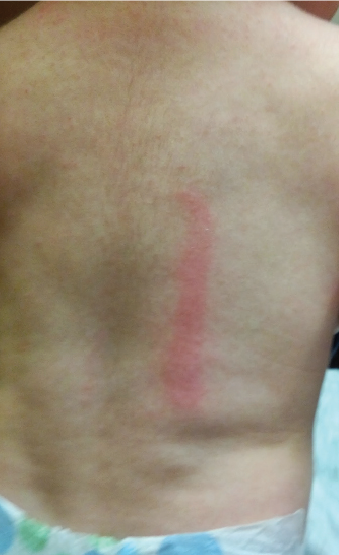
Examination revealed a normal, well-appearing child with a mild erythematous, macular rash in a collar-like fashion around his neck. Using a tongue depressor with light pressure to draw the letter H on the patient’s back produced a large, expanding, erythematous flare and linear wheal in the same shape.
Based on the patient’s clinical signs and symptoms, and that a wheal and flare reaction occurred with applied pressure from a tongue depressor, he received a diagnosis of simple dermographism.
Dermographism, the most common type of physical urticaria, can be divided into 2 general categories, simple and symptomatic.1,2 Simple dermographism involves inflammation and swelling of the skin, whereas symptomatic dermographism also includes itching.1-5 The wheal and flare reaction usually develops in a few minutes but also can develop many hours after the initial application of mechanical pressure.2 In general, the urticarial reaction lasts approximately 30 minutes but has been known to be prolonged for 1 to 2 days.2
The differential diagnosis for simple dermographism includes symptomatic dermographism, contact dermatitis, drug-induced dermatitis, viral exanthems, mastocytosis, and rashes with thyroid or endocrine etiologies.2
While the exact prevalence of simple dermographism is not known in the pediatric population, studies have estimated it at 10% to 24%, with a predominance in girls; the prevalence in the general population is estimated at 1.5% to 5%.2,3,6
The pathophysiology of simple dermographism is unknown; however, it is theorized that mechanical trauma causes a release of an unknown antigen that then interacts with IgE-mediated receptors on mast cells.2,3 This interaction causes a release of histamine from mast cells, which increases both venous and arteriolar permeability, creating the classic wheal and flare reaction.2,3
 A thorough history and physical are important in the diagnosis of simple dermographism, especially a history of the use of any medications, since dermographism has been associated with the use of penicillin, famotidine, and atorvastatin.2,3
While standardized and objective methods of diagnosing simple dermographism using instruments that measure applied pressure are available, administering mild pressure to the skin using a tongue blade or closed ballpoint pen will produce the pathognomic reaction in the primary care setting.1,2 Although laboratory tests usually are not required to make the diagnosis of simple dermographism, a biopsy of the rash might be useful to rule out other disorders.2
Management is straightforward and focuses on improvement of symptoms. Nonpharmacologic treatment of dermographism includes identifying and limiting the physical triggers, such as the use of loose-fitting clothes.2 Some studies have suggested that stress can be a trigger; thus, avoidance of those psychological factors is important, as well.2,3,7 The main pharmacologic treatment is a nonsedating histamine H1-receptor antagonist such as cetirizine, although the evidence of effectiveness is inconsistent.2-5 Dermographism generally is a self-limiting disease that often resolves in 5 years.2
After the mother was reassured that her infant son’s rash was a self-limiting, benign condition, she chose not to use any pharmacologic treatment for her child, instead opting for loose-fitting clothing.
REFERENCES
1. Mlynek A, Vieira dos Santos R, Ardelean E, et al. A novel, simple, validated and reproducible instrument for assessing provocation threshold levels in patients with symptomatic dermographism. Clin Exp Dermatol. 2013;38(4):360-366.
2. Mecoli CA, Morgan AJ, Schwartz RA. Symptomatic dermatographism: current concepts in clinical practice with an emphasis on the pediatric population. Cutis. 2011;87(5):221-225.
3Taşkapan O, Harmanyeri Y. Evaluation of patients with symptomatic dermographism. J Eur Acad Dermatol Venereol. 2006;20(1):58-62.
4. Magerl M, Schmolke J, Metz M, Zuberbier T, Siebenhaar F, Maurer M. Prevention of signs and symptoms of dermographic urticaria by single-dose ebastine 20 mg. Clin Exp Dermatol. 2009;34(5):e137-e140.
5. Toda S, Takahagi S, Mihara S, Hide M. Six cases of antihistamine-resistant dermographic urticaria treated with oral ciclosporin. Allergol Int. 2011; 60(4):547-550.
6. Martorell A, Sanz J, Ortiz M, et al. Prevalence of dermographism in children. J Investig Allergol Clin Immunol. 2000;10(3):166-169.
7. Wallengren J, Isaksson A. Urticarial dermographism: clinical features and response to psychosocial stress. Acta Derm Venereol. 2007;87(6):493-498.