
Hair Breakage and Spontaneous Hair Loss in 2 Sailors Aboard an Aircraft Carrier
While aboard a US Navy aircraft carrier in the North Arabian Gulf, 2 active-duty sailors presented independently to the medical department with remarkably similar symptoms of hair strand breakage while brushing, along with spontaneous hair loss.
The first patient was a 33-year-old woman who presented with a 1-month history of these symptoms, along with finding a large amount of hair in her helmet at the end of each day. The patient worked primarily on the flight deck in high heat and humidity. The second patient was a 43-year-old woman who worked in a different part of the ship and presented with nearly identical symptoms 2 weeks after the first patient.
Aboard the ship, both patients lived in a tight berthing area shared by 213 women; coincidentally, they slept 1.5 m from each other. Both women denied using the other’s hairbrush, hair ties, or any hair products.

On physical examination of the first patient, numerous cream-colored nodules measuring 0.3 to 0.5 mm were seen on the hair shafts (Figure 1). The nodules were irregular, shiny, and gelatinous-appearing, with even distribution on the hair shafts proximally to distally. The hair strands would break with tension at the areas of the nodules. Examination findings of the scalp were unremarkable.
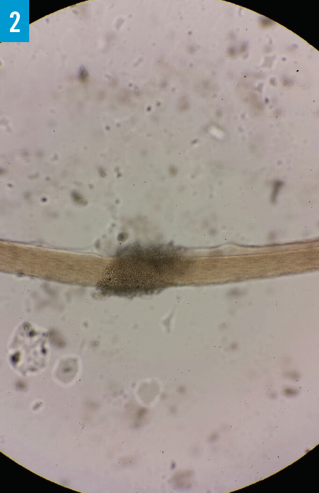
Microscopic examination of a potassium hydroxide preparation test specimen showed that the nodules were made up of a mass of small ovals that surrounded and invaded the hair shaft (Figure 2).

What is responsible for the women’s symptoms?
- Pediculosis capitis
- White piedra
- Trichorrhexis nodosa
- Monilethrix
- Peripilar keratin casts
Answer and podcast on the next page.
Answer: White Piedra
While aboard a US Navy aircraft carrier in the North Arabian Gulf, 2 active-duty sailors presented independently to the medical department with remarkably similar symptoms of hair strand breakage while brushing, along with spontaneous hair loss.
The first patient was a 33-year-old woman who presented with a 1-month history of these symptoms, along with finding a large amount of hair in her helmet at the end of each day. The patient worked primarily on the flight deck in high heat and humidity. The second patient was a 43-year-old woman who worked in a different part of the ship and presented with nearly identical symptoms 2 weeks after the first patient.
Aboard the ship, both patients lived in a tight berthing area shared by 213 women; coincidentally, they slept 5 feet from each other. Both women denied using the other’s hairbrush, hair ties, or any hair products.
On physical examination of the first patient, numerous cream-colored nodules measuring 0.3 to 0.5 mm were seen on the hair shafts (Figure 1). The nodules were irregular, shiny, and gelatinous-appearing, with even distribution on the hair shafts proximally to distally. The hair strands would break with tension at the areas of the nodules. Examination findings of the scalp were unremarkable.

Microscopic examination of a potassium hydroxide (KOH) preparation test specimen showed that the nodules were made up of a mass of small ovals that surround and invaded the hair shaft (Figure 2).
Based on the clinical presentation and test results, both patients received a diagnosis of white piedra.
Shaving of the head, which usually is the first-line treatment for white piedra, was avoided in order to preserve the morale of the 2 women sailors and to avoid possible panic in a 213-woman berthing area. The patients were prescribed a 10-week regimen of oral itraconazole, 100 mg/d, and topical fluconazole shampoo, 2%. After 4 weeks of treatment, the proximal 6 cm of the women’s hair strands were clear of the nodules, and the remaining distal nodules had decreased in size. At 8 weeks of treatment, all symptoms had resolved. Both patients completed 10 weeks of treatment and were monitored for 3 months with no return of symptoms.
Discussion
White piedra, or trichomycosis nodularis, is a common superficial fungal infection of the hair shaft caused by Trichosporon asahii, formerly known as Trichosporon beigelii.1,2 Trichosporon forms soft white to light brown nodules that are loosely adherent to the hair shaft. Cuticular invasion occurs within the nodules, weakening the shaft’s structural integrity and leading to breakage.
White piedra is normally found in the hair of the genitals, beard, moustache, eyebrows, eyelashes, axillae, and rarely the scalp.1-3 This is in contrast to black piedra, caused by Piedraia hortae, which is characterized by the presence of firmly attached, black/brown, hard nodules that are more commonly attached to scalp hair. White piedra is believed to be sporadic and prepathologic; Trichosporon is carried asymptomatically on the hair and skin of the patient.3,4
White piedra is more common in temperate and semitropical climates, including the Southern United States, and can be found in soil and vegetable matter or on body surfaces.4,5 People of all ages are affected, and age and sex incidence varies from country to country.2 The specific mode of transmission is not clear, and most suggest infection occurs from soil or animals; however, person-to-person transmission has been suggested as possible but rare.1-3,6
Patients with white piedra normally seek medical care because of hair loss, or they find visible nodules on their hair. The diagnosis of white piedra can be made purely by the visualization of the characteristic white to light brown soft nodules seen on the shafts of affected hairs.1,2,6 Microscopic examination of these nodules with 10% KOH preparation shows septate hyphae with arthroconidia. The hair shafts will have a positive pull test, breaking at the location of the nodules when tension is applied.1-3,5
Given that white piedra can be easily misdiagnosed, the differential diagnosis is important and includes pediculosis capitis, trichomycosis axillaris, peripilar keratin casts, monilethrix, and trichorrhexis nodosa. Wood light examination, KOH preparation testing, and fungal culture testing help in differentiating these conditions.1,2,7
Treatment of superficial infection of white piedra is best achieved by shaving off all of the hair in the affected areas and the use of a topical antifungal to the scalp. Ketoconazole shampoo is most commonly used for a period of several months. The specific treatment duration depends on the treatment response.1,2,5,6 However, if the patient refuses to shave his or her hair, oral daily itraconazole, 100 mg, is suggested in addition to the topical regimen.1,3,5 Treatment can be a therapeutic challenge, since several topical and systemic antifungal agents may not eliminate the infection completely.1,6
White piedra is one of many interesting and rare challenges that present in operational medicine. This case may represent a rare instance of person-to-person transmission of the pathologic organism, possibility stemming from the harsh conditions while living on a deployed aircraft carrier. Additionally, because both patients did not desire to shave their hair off as part of treatment, both were successfully treated with 10 weeks of oral and topical antifungals. This further supports the viability of this alternative less-aggressive treatment.
Robert J. Long, MD, is a lieutenant and flight surgeon in the US Navy Medical Corps at Beaufort Marine Corps Air Station in Beaufort, South Carolina.
Disclosure:
The views expressed herein are those of the author and do not necessarily reflect the official policy or position of the Department of the Navy, the Department of Defense, or the United States Government.
References:
- Hay RJ, Moore MK. Mycology. In: Burns T, Breathnach S, Cox N, Griffiths C, eds. Rook’s Textbook of Dermatology. Vol 2. 7th ed. Malden, MA: Blackwell Publishing Inc; 2004:chap 31.
- Tambe SA, Dhurat SR, Kumar CA, et al. Two cases of scalp white piedra caused by Trichosporon ovoides. Indian J Dermatol Venereol Leprol. 2009;75(3):293-295.
- Kalter DC, Tschen JA, Cernoch PL, et al. Genital white piedra: epidemiology, microbiology, and therapy. J Am Acad Dermatol. 1986;14(6):982-993.
- Ellner K, McBride ME, Rosen T, Berman D. Prevalence of Trichosporon beigelii: colonization of normal perigenital skin. J Med Vet Mycol. 1991;29(2):99-103.
- Guidelines of care for superficial mycotic infections of the skin: Piedra. Guidelines/Outcomes Committee. American Academy of Dermatology. J Am Acad Dermatol. 1996;34(1):122-124.
- Khandpur S, Reddy BS. Itraconazole therapy for white piedra affecting scalp hair. J Am Acad Dermatol. 2002;47(3):415-418.
- Gold I, Sommer B, Urson S, Schewach-Millet M. White piedra: a frequently misdiagnosed infection of hair. Int J Dermatol. 1984;23(9):621-623.