
Elephantiasis Nostras Verrucosa
A 53-year-old man presented to the emergency department with a cellulitis infection of his right lower extremity. This was his 15th admission in the last 2 years for the same complication. The patient had a longstanding history of Child-Pugh class C cirrhosis secondary to chronic hepatitis C infection and a history of alcohol abuse. Although he had been prescribed diuretics to reduce his ascites and lower-extremity edema, the patient did not take his medications regularly and was not amenable to wearing compression stockings.
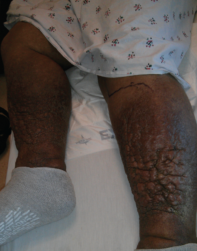
Physical examination. On examination, there were significant verrucous, fibrotic changes to the skin extending from below the knees to the feet (Figures). The skin was firm to the touch and malodorous. There was significant hemosiderin deposition. The pedal pulses were easily palpable. Skin examination revealed a positive Stemmer sign bilaterally.
Discussion. Elephantiasis nostras verrucosa (ENV) classically occurs in the setting of chronic nonfilarial lymphedema. Prolonged lymphostasis of the affected extremity eventually leads to accumulation of protein within the interstitial fluid, which in turn induces proliferation of fibroblasts. It is believed that these fibroblasts cause the progressive fibrosis of the dermis and subcutaneous tissue that is characteristic of this condition.1
Recurrent soft tissue infections such as cellulitis and erysipelas often are the underlying cause of the lymphedema. Repetitive lymphangitis can cause fibrosis of the lymphatic drainage system, leading to obstruction.1 Other causes include occult malignancy, particularly lymphoma; iatrogenic causes such as surgery or radiation therapy; and scleroderma. Obesity and chronic venous insufficiency are significant risk factors.2
As the condition progresses, bacteria begin to colonize the abnormal skin, causing a fetid odor. Ulcerations can develop with skin breakdown. These developments in turn lead to more local infections, which go on to drive the cycle of disease progression further.
Management. Eradication of ENV skin changes can be challenging. Identifying and correcting the underlying cause of the lymphedema is the most important factor in management. Strategies to reduce or reverse skin changes include compression stockings, adjunctive use of systemic or topical retinoids, and surgical debridement. Keratinolytics and deodorant powders also can be used.3 Response to treatment has largely been described in case reports; there is a paucity of large-scale studies describing treatment outcomes.

References:
- Sisto K, Khachemoune A. Elephantiasis nostras verrucosa: a review. Am J Clin Dermatol. 2008;9(3):141-146.
- Dean SM, Zirwas MJ, Horst AV. Elephantiasis nostras verrucosa: an institutional analysis of 21 cases. J Am Acad Dermatol. 2011;64(6):1104-1110.
- Fredman R, Tenenhaus M. Elephantiasis nostras verrucosa. Eplasty. 2012;12:ic14.
1. Braverman IM. Skin Signs of Systemic Disease. 2nd ed. Philadelphia: WB Saunders Co; 1981:809-813.
2. Moschella SL, Hurley HJ, eds. Dermatology. 3rd ed. Philadelphia: WB Saunders Co; 1992:729,1165.
3. Odom RB, James WD, Berger TG, eds. Andrews' Diseases of the Skin: Clinical Dermatology. 9th ed. Philadelphia: WB Saunders Co; 2000:319-320.
4. Erickson QL, Kobayashi T, Vogel PS. Photo quiz. Woody edema of the legs. Am Fam Physician. 2003; 67:583-584.
5. Davis PL, Paoli P. Elephantiasis nostra verrucosa: lymphostatic verrucosis. Arch Dermatol. 1955;71:644.