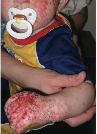
Eczema Herpeticum: A Boy With Multiple Monomorphic Erosions
A 2-year-old boy presented with a mild fever, malaise, and an extensive rash of 4-days’ duration on the face, dorsum of the left hand, and left forearm. The rash began as pruritic, monomorphic, dome-shaped vesicles that quickly became crusted and eroded. The boy had a history of atopic dermatitis since 8 months of age, and his mother had a cold sore on her lip 2 weeks prior to the boy’s presentation.
On examination, the child’s temperature was 37.8°C, his heart rate was 88 beats per minute, and his respiratory rate was 22 breaths per minute. There were multiple, small, round, grouped, punched-out monomorphic erosions with crusting and an erythematous base on the face, dorsum of the left hand, and left forearm. Some of the lesions coalesced into confluent areas of denudation. Shotty lymph nodes were noted in the cervical areas. The rest of the physical examination results were unremarkable.
Based on the clinical appearance, a diagnosis of eczema herpeticum was made. The diagnosis was confirmed by a Tzanck test that showed multinucleated giant cells and a direct florescent antibody test that was positive for herpes simplex virus (HSV) type 1.
The patient was treated with intravenous acyclovir and proper wound care. Viral culture from an opened vesicle grew HSV type 1 on day 5 of therapy, and by that time the lesions had significantly improved.
ECZEMA HERPETICUM: AN OVERVIEW
Eczema herpeticum refers to an extensive disseminated cutaneous infection with HSV in patients with atopic dermatitis.1 The condition was first described in 1887 by Moritz Kaposi.2 Some authors use the terms “eczema herpeticum” and “Kaposi varicelliform eruption” interchangeably. However, most authors use the term “Kaposi varicelliform eruption” to describe a similar clinical picture without specifying the causative virus (eg, HSV, coxsackievirus A16, and vaccinia) in patients with variable skin conditions. These skin conditions can include atopic dermatitis, irritant contact dermatitis, seborrheic dermatitis, ichthyosis vulgaris, psoriasis, burns, pemphigus vulgaris, pityriasis rubra pilaris, dyskeratosis follicularis, congenital ichthyosiform erythroderma, mycosis fungoides, Wiskott-Aldrich syndrome, Sézary syndrome, Darier disease, and Hailey-Hailey disease).3
Epidemiology
The true incidence of eczema herpeticum is not known. It is estimated that approximately 1.4% to 3% of patients with atopic dermatitis exposed to HSV develop eczema herpeticum.4,5 The condition is more common in children than in adults, presumably because of its relationship to atopic dermatitis.6,7 Both sexes are affected equally,7 most cases are sporadic,6 and there is no seasonal variation.7
Etiopathogenesis
Eczema herpeticum is most commonly caused by primary or secondary/recurrent infection with HSV-1 and, less commonly, HSV-2.8 Transmission is by direct contact with an infected individual, autoinoculation, or reactivation of latent HSV.7
Individuals with atopic dermatitis have impairment of the barrier function of the skin and decreased local cell-mediated and humoral immune responses that make them susceptible to HSV. In patients with atopic dermatitis, the stratum corneum can be dysfunctional as a result of defects in filaggrin, reduced content of ceramides, and physical trauma.9 Tight junctions reside immediately below the stratum corneum and regulate the selective permeability of the paracellular pathway. Patients with atopic dermatitis have reduced levels of claudin-1, a key tight junction adhesive protein.9 In addition, patients with atopic dermatitis have a decreased ability to produce cathelicidin, β-defensins, and interferon. Their ability to recruit plasmacytoid dendritic cells into the lesional skin is also impaired. All these factors make the lesional skin susceptible to viral infections.3,4
Risk factors for eczema herpeticum include early onset of atopic dermatitis, more severe atopic dermatitis, associated allergic diseases (eg, asthma, allergic rhinitis, food allergy), higher serum immunoglobulin E levels, higher circulating eosinophil counts, and immunosuppression.7,9,10
Histopathology
Histologic findings include intranuclear inclusions in the basal layer of the epidermis; keratinocytes undergoing balloon degeneration, multinucleation, and acantholysis; and epithelial necrosis and ulceration.8

Clinical Manifestations
Typically, eczema herpeticum presents as clusters of dome-shaped monomorphic umbilicated vesicles in an area affected by atopic dermatitis.10,11 The vesicles spread, and they soon become vesiculopustular, hemorrhagic, crusted, and eroded.10 The punched-out erosions are surrounded by erythema and are painful,10 and they may coalesce to form denuded areas.11,12 New crops may continue for 7 to 10 days. Characteristically, the crops are at the same stage of evolution.7 Sites of predilection include the head, neck, trunk, and upper extremities.11,13,14 Fever, malaise, and regional lymphadenopathy are often present.14
The severity of eczema herpeticum varies from mild transient disease to a fulminating fatal disorder involving the visceral organs. Eczema herpeticum caused by the primary infection is usually more severe than the recurrent one.
Diagnosis
The diagnosis is mainly clinical, based on the patient history and physical examination findings. Eczema herpeticum should be suspected if there is a sudden deterioration of atopic dermatitis with grouped and locally disseminated monomorphic vesicles with subsequent painful punched-out erosions.10 A positive Tzanck smear showing epithelial multinucleated giant cells is highly suggestive. The diagnosis can be confirmed by direct fluorescent antibody staining of vesicular fluid, polymerase chain reaction for viral DNA, and viral culture.13
Differential Diagnosis
Differential diagnosis includes chickenpox, herpes zoster, impetigo, cellulitis, contact dermatitis, Stevens-Johnson syndrome, pustular psoriasis, pemphigus vulgaris, bullous pemphigoid, bullous lupus erythematosus, Henoch-Schönlein purpura, and eczema vaccinatum.8
Complications
The most common complication is secondary bacterial infection, primarily with Staphylococcus aureus and Streptococcus pyogenes. Scarring and postinflammatory hyper/hypo pigmentation are uncommon but may occur.7 Other complications include blepharitis, keratoconjunctivitis, and viremia. The latter can result in multiorgan involvement such as hepatitis, meningitis, and encephalitis.7,9 Viremia in pregnancy may lead to fetal demise, miscarriage, and neonatal herpes infection.15
Prognosis
Untreated, the mortality rate for eczema herpeticum can be as high as 10%, mainly as a result of systemic dissemination of the viral infection and septicemia.7,8 Nevertheless, the majority of cases are mild and localized. With proper treatment, lesions usually heal within 2 to 6 weeks.12,13
Management
Oral acyclovir is the drug of choice for healthy, immunocompetent children with mild disease. Intravenous acyclovir is recommended for children who have severe disease, who have systemic involvement, or who are immunocompromised.7
Vidarabine, trifluridine, cidofovir, and foscarnet can be considered in resistant cases.16 Antibiotic therapy is warranted if bacterial superinfection is suspected.13 Topical corticosteroids or calcineurin inhibitors for residual adjacent eczema are not generally recommended in the acute phase of the disease, but they may be considered once systemic antiviral therapy has been started and the disease is under control.14
Alexander K. C. Leung, MD, is a Clinical Professor of Pediatrics at the University of Calgary and a pediatric consultant at the Alberta Children’s Hospital in Calgary, Alberta, Canada.
Benjamin Barankin, MD, is a dermatologist and the Medical Director and Founder of the Toronto Dermatology Centre in Toronto, Ontario, Canada.
REFERENCES
1. Takahashi R, Sato Y, Kurata M, Yamazaki Y, Kimishima M, Shiohara T. Pathological role of regulatory T cells in the initiation and maintenance of eczema herpeticum lesions. J Immunol. 2014;192(3):969-978.
2. Kaposi M. Pathologie und Therapie der Hautkrankheiten. Vienna, Austria: Urban and Schwarzenberg; 1887.
3. Shenoy R, Mostow E, Cain G. Eczema herpeticum in a wrestler. Clin J Sport Med. 2015;25(1):e18-e19.
4. Leung DY. Why is eczema herpeticum unexpectedly rare? Antiviral Res. 2013;98(2):153-157.
5. Tay YK, Khoo BP, Goh CL. The epidemiology of atopic dermatitis at a tertiary referral skin center in Singapore. Asian Pac J Allergy Immunol. 1999;17(3):137-141.
6. Ferrari B, Taliercio V, Luna P, Abad ME, Larralde M. Kaposi’s varicelliform eruption: a case series. Indian Dermatol Online J. 2015;6(6):399-402.
7. Khan A, Shaw L, Bernatoniene J. Fifteen-minute consultation: eczema herpeticum in a child. Arch Dis Child Educ Pract Ed. 2015;100(2):64-68.
8. Mackool BT, Goverman J, Nazarian PM. Case records of the Massachusetts General Hospital. Case 14-2012: a 43-year-old woman with fever and a generalized rash. N Engl J Med. 2012;366(19):1825-1834.
9. Leung AK, Hon KL. Atopic Dermatitis: A Review for the Primary Care Physician. New York, NY: Nova Science Publishers, Inc; 2012:1-113.
10.Liaw FY, Huang CF, Hsueh JT, Chiang CP. Eczema herpeticum: a medical emergency. Can Fam Physician. 2012;58(12):1358-1361.
11. Vora RV, Pilani AP, Jivani NB, Kota RK. Kaposi varicelliform eruption. Indian Dermatol Online J. 2015;6(5):364-366.
12. Blanter M, Vickers J, Russo M, Safai B. Eczema herpeticum: would you know it if you saw it? Pediatr Emerg Care. 2015;31(8):586-588.
13. Luca NJC, Lara-Corrales I, Pope E. Eczema herpeticum in children: clinical features and factors predictive of hospitalization. J Pediatr. 2012;161(4):671-675.
14. Wollenberg A. Eczema herpeticum. Chem Immunol Allergy. 2012;96:89-95.
15. DiCarlo A, Amon E, Gardner M, Barr S, Ott K. Eczema herpeticum in pregnancy and neonatal herpes infection. Obstet Gynecol. 2008;112(2 Pt 2):455-457.
16. Frisch S, Siegfried EC. The clinical spectrum and therapeutic challenge of eczema herpeticum. Pediatr Dermatol. 2011;28(1):46-52.


