Do You Know Your Nevi?

Case 1:
The square, flat, red lesion on the left lower lip vermilion of an 11-day-old boy developed about 24 hours after birth. Because the lesion seemed to have increased in size, the mother was anxious about it, and a dermatological consultation was obtained.
What is your clinical impression?
(Answer on next page.)
ANSWER—Case 1: Strawberry nevus

Strawberry nevi are developmental abnormalities consisting of dilated vessels in the epidermis surrounded by masses of proliferating endothelial cells. Synonyms include superficial capillary hemangioma, juvenile hemangioma, and benign hemangioendothelioma of childhood.
The lesions are usually not present at birth but occur within the first few days or weeks of life in 1% to 3% of births.1 There is a female predominance of 3:1. Strawberry nevi are often singular. They usually proliferate for 8 to 18 months then slowly spontaneously involute over the next few years with little to no disfigurement.
Most strawberry nevi require no treatment. The few lesions with functional impairment, deep ulceration, or infection are treated, as are those thought to be a cosmetic concern by the patient or parent. Treatments include oral corticosteroids, intralesional corticosteroids, interferon alpha-2a, and pulsed dye laser therapy.
This infant’s lesion was treated with intralesional corticosteroid injections and flashlamp pulsed dye laser on several occasions at about monthly intervals and had regressed.
REFERENCES:
1. Habif TP. Clinical Dermatology: A Color Guide to Diagnosis and Therapy. 3rd ed. St Louis: Mosby; 1996:722-723.
2. Weedon D. Skin Pathology. London: Churchill Livingstone: 1997:827-828.
(Next case on next page.)
 Case 2:
This oblong, 4 3 3-mm, dark reddish brown, slightly elevated lesion had been present on an 8-year-old girl’s right mid back for about 2 years. The lesion had gradually enlarged and had become much darker over the past 6 months. It was asymptomatic. Her father and one brother had a history of malignant melanoma.
Is it likely that this lesion is malignant?
(Answer on next page.)
ANSWER—Case 2: Spitz nevus

The lesion was excised in the office. Pathologic diagnosis was Spitz nevus—an uncommon, benign melanocytic neoplasm. Microscopically, the lesion exhibited most of the major criteria of Spitz nevus, including asymmetry, spindle cells, cellular maturation, and absence of pagetoid spread of single melanocytes. A few minor criteria were also present, including perivascular inflammation, absence of nuclear pleomorphism, no deep atypical mitosis, deep outlying solitary nevus cells, and superficial edema and telangiectasia.1
Spitz nevus has been termed spindle cell nevus, epithelioid cell nevus, nevus of large spindle and/or epithelioid cells, and benign juvenile melanoma. It is usually a solitary lesion and may occur on any part of the body. The nevus appears suddenly—the patient may date its onset. It is most common in children. Spitz nevi have been reported in 0.5% to 1.0% of all excised nevi in children and adolescents.2 Recurrence rate is low even after incomplete excision.3
The typical Spitz nevus is a pink or flesh-colored papule or nodule, although pigmented variants are not uncommon. Other variants include atypical, halo, and malignant-type. Histologic examination is mandatory to differentiate the lesion from malignant melanoma; however, even experts in dermatopathology may have difficulty in distinguishing an atypical Spitz nevus from melanoma.4 Numerous histologic and immunochemical tests may be used in the differentiation of these lesions.1
Treatment of Spitz nevi is by complete excision.
REFERENCES:
1. Weedon D. Skin Pathology. 2nd ed. London: Churchill Livingstone: 2002; 811-814.
2. Weedon D. The Spitz nevus. Clin Oncol. 1984;3:493-507.
3. Kaye VN, Dehner LP. Spindle and epithelioid cell nevus (Spitz nevus). Natural history following biopsy. Arch Dermatol. 1990;126(12):1581-1583.
4. Barnhill RL, Argenyi ZB, From L, et al. Atypical Spitz nevi/tumors: lack of consensus for diagnosis, discrimination from melanoma, and outcome. Hum Pathol. 1999;30(5):513-520.
(Next case on next page.)
 Case 3:
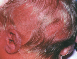
The salmon-colored, hairless, circumscribed lesions in the left parietal and occipital areas of a female newborn’s scalp were noted at birth.
Is biopsy warranted?
(Answer on page 78.)
ANSWER—Case 3: Nevus sebaceus of Jadassohn

The infant was referred to a pediatric dermatologist who diagnosed nevus sebaceus of Jadassohn by the clinical appearance of the lesion. An incisional biopsy was recommended for confirmation but was not done.
Nevus sebaceus of Jadassohn presents as well-circumscribed, linear or oval, hairless plaques mainly on the face and scalp. The lesion may be a few millimeters to several centimeters in diameter and is usually present at birth, although it may present in early childhood and rarely in adults. It is benign, often sporadic and solitary.
Histologically, nevus sebaceus of Jadassohn (organoid nevus) is a complex hamartoma that involves the pilosebaceous follicle, the epidermis, and often other adnexal structures.
Multiple sebaceous nevi may be associated with cerebral, ocular, vascular, urogenital, or skeletal abnormalities as part of the epidermal nevus syndrome, also known as Schimmelpenning syndrome.l Patients with neurological signs or symptoms and those with large lesions in the centrofacial area should be considered for neurological assessment, including cerebral imaging.2
Some lesions are hyperpigmented and may appear to be a verrucous epidermal nevus. The sebaceous gland secretions produce a yellow coloration, which is less prominent after infancy. After puberty with hormonal stimulation, the lesions become “greasy” and thicken and may have the appearance of verruca vulgaris.
Of the tumors associated with nevus sebaceus of Jadassohn, basal cell carcinoma has been estimated to occur in 6.5% to 50% of lesions.3 However, more recent studies estimate the development of basal cell carcinoma to be quite low.4,5 Trichoblastoma, which mimics basal cell carcinoma and is benign, appears to be quite common with a strong female predominance. Other associated neoplasms are syringocystadenoma papilliferum (the most common secondary benign neoplasm), apocrine cystadenoma and carcinoma, spiradenoma, keratoacanthoma, leiomyoma, piloleiomyoma, squamous cell carcinoma, and malignant eccrine poroma.4,6,7
Because of concern for malignancy development, which is heralded by nodule formation, surgical excision has been the traditional treatment recommendation. However, prophylactic surgery is probably unnecessary in most children. Most neoplasms that develop are benign and occur in adults older than 40 years.8 The malignancies that do develop are usually low-grade, and rates of metastases of these tumors are low.5 In select cases, plastic surgery may be a consideration. Electrosurgery and cryotherapy may lead to tumor recurrence.
REFERENCES:
1. Happle R. Epidermal nevus syndromes [published correction appears in Semin Dermatol. 1995;14(3):259]. Semin Dermatol. 1995;14(2):111-121.
2. Davies D, Rogers M. Review of neurological manifestations in 196 patients with sebaceous naevi. Australas J Dermatol. 2002;43(1):20-23.
3. Barankin B, Shum D, Guenther L. Tumors arising in nevus sebaceus: a study of 596 cases. J Am Acad Dermatol. 2001;45(5):792-793; author reply 794.
4. Kaddu S, Schaeppi H, Kerl H, Soyer HP. Basaloid neoplasms in nevus sebaceus. J Cutan Pathol. 2000;27(7):327-337.
5. Cribier B, Scrivener Y, Grosshans E. Tumors arising in nevus sebaceus: a study of 596 cases. J Am Acad Dermatol. 2000;42(2, pt 1):263-268.
6. Chun K, Vázquez M, Sánchez JL. Nevus sebaceus: clinical outcome and considerations for prophylactic excision. Int J Dermatol. 1995;34(8):538-541.
7. Shapiro M. Johnson V Jr, Witmer W, Elenitsas R. Spiradenoma arising in a nevus sebaceus of Jadassohn: case report and literature review. Am J Dermatopathol.1999;21(5):462-467.
8. Habif TP. Clinical Dermatology: A Color Guide to Diagnosis and Therapy. 3rd ed. St Louis: Mosby; 1996:715.
(Next case on next page.)
 Case 4:
A 12-year-old girl presented with her caregiver who was concerned about a dark brown, slightly elevated lesion in the girl’s postauricular area. The lesion was about 0.5 cm in diameter and was surrounded by a ring of light tan, finely eczematoid skin. It had been present for years and was asymptomatic. Because the lesion had grown over time, the caregiver wanted it removed.
What would you include in the differential for this lesion?
(Answer on next page.)
ANSWER—Case 4: Pigmented compound nevus
 In the differential diagnosis of this particular nevus are halo nevus and Meyerson nevus. A halo nevus is surrounded by a white halo of depigmentation, as the name implies, and occurs in association with a lymphocytic infiltration and destruction of nevus cells. A Meyerson nevus is a pigmented lesion with an eczematoid halo that may be accompanied by pruritus. In contrast to the halo nevus, the Meyerson nevus is not associated with destruction of nevus cells.
From the gross appearance of this patient’s lesion, it was felt that it was a Meyerson nevus; however, microscopic examination of the excised lesion revealed findings consistent with a pigmented compound nevus, with tailing off of the nevus cells around the periphery. The nevus lacked epidermal spongiosis required for diagnosis of a Meyerson nevus.
Common melanocytic nevi are classified as junctional, compound, dermal, or combined according to the location of the nevus cells in the skin. Compound nevi are located at the dermoepidermal junction and upper dermis. These moles are slightly elevated and may be flesh-colored or shades of brown with or without hair.
Most compound nevi are benign and do not require excision. However, they may be removed for cosmesis, when malignancy is a concern, or when the mole causes irritation.
(Next case on next page.)

Case 5:
This flesh-colored, lobulated, 0.5-cm nodule with several hairs protruding through its surface on a 16-year-old girl’s occipital scalp had been present since birth and had recently begun to enlarge.
Can you identify this lesion?
(Answer on page 79.)
ANSWER—Case 5: Intradermal nevus

This patient has a lobulated amelanotic intradermal nevus. Intradermal nevi are the most common type of melanocytic nevi and are benign. Most of these lesions occur in adults, are usually tan or brown, elevated, and larger than compound or junctional nevi. Melanin production by nevus cells decreases with increasing depth of location of the nevus.1
As per the patient’s request, the lesion was removed. This was done by shave excision under local anesthesia in the office. The base of the lesion was electrocoagulated and curetted. Pathologic examination revealed it to be an intradermal nevus.
REFERENCE:
1. Sams WM Jr, Lynch PJ, eds. Principles and Practice of Dermatology. 2nd ed. New York: Churchill Livingstone; 1996:258.


