
Diffuse Extragenital Lichen Sclerosus
An 84-year-old woman presented with worsening pain and burning of the skin on her neck and torso that had begun several months prior. She reported that she had been diagnosed with lichen sclerosus (LS) approximately 12 years ago. She stated that she initially had a small, silver dollar–sized area on her right breast that had slowly progressed to multiple lesions and had begun to cover her neck, back, and chest. She could not recall the specific different treatment regimens she had tried.
Physical examination. On physical examination, she exhibited diffuse, porcelain white to pink, sclerotic, and atrophic papules and plaques distributed on her neck, upper torso, breasts, and thighs, with noticeable sparing of her genital area (Figures 1-5).


Figures 1-5. Diffusely scattered, porcelain white to pink, sclerotic, and atrophic papules and plaques on the chest, back, and extremities of an 84-year-old woman.
Diagnostic tests. A 60-mm punch biopsy was taken of a larger sclerotic plaque at the inferior chest, at the location of the xiphoid process, to rule out various panniculitides (Figure 6). Histologic examination revealed epidermal atrophy, hyperkeratosis, basal vacuolar changes, and sclerosis and edema within the dermis (Figure 7), findings consistent with LS.
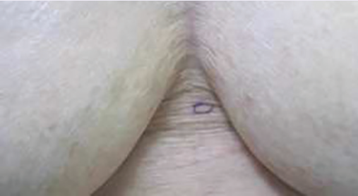
Figure 6. Sclerotic and atrophic papule on sternum marked for punch biopsy.

Figure 7. Medium-power view of the punch biopsy specimen showing epidermal atrophy, hyperkeratosis, basal vacuolar changes, and sclerosis and edema within the papillary dermis.
Treatment. The patient was provided with topical triamcinolone to apply to the affected areas and was referred to a rheumatologist for evaluation of any other possible underlying autoimmune process, given the extensive involvement. Other systemic treatment options were discussed with the patient. Nevertheless, the patient declined systemic treatment and further rheumatologic evaluation.
Outcome of the case. A follow-up phone call to the patient revealed that she had experienced some mild improvement with topical triamcinolone, but she declined any further follow-up.
Discussion. The term lichen sclerosus was coined in 1887, and the condition has been synonymously called kraurosis vulvae or vulvar dystrophy when affecting female genitalia and balanitis xerotica obliterans when affecting male genitalia.1 These and other names, including guttate scleroderma and lichen sclerosus et atrophicus, have been replaced simply with the term lichen sclerosus for both the genital and nongenital conditions.1 LS is considered as a relatively uncommon disease, with extragenital presentations even less common. In fact, only 6% of LS cases are solely extragenital.1
This condition most commonly affects women during their 40s or 50s.2 Children also can be affected, although much less commonly.2 Adults experience a more chronic disease process, while children usually have self-limiting cases.3
The exact cause of LS remains unknown. Some researchers have demonstrated the presence of a specific T-cell response and autoantibody formation to extracellular matrix protein 1. This protein resides in the epidermis and dermis and plays a key role in epidermal and keratinocyte differentiation, dermal protein interactions, dermal structure organization, regulating assembly of collagen fibrils, and aiding in growth-factor binding. Immunoglobulin G autoantibodies have been found in 74% of women with genital LS cases. Some authors believe that such a finding may be a secondary phenomenon.1,4,5 Additional cases of predominantly vulvar bullous LS have exhibited a significant amount of autoantibody formation to basement membrane zone proteins BPAG1 and BPAG2.1
Additionally, LS has been associated with trauma; HLA class II antigens DQ7, DQ8, and DQ9; and infection with Borrelia burgdorferi, Epstein-Barr virus, and hepatitis C.1 Autoimmune processes have strong associations, as well, reportedly coinciding in up to 60% of affected women.6 Other associated conditions include thyroid disease, pernicious anemia, diabetes mellitus, vitiligo, and alopecia areata.4,5 There is an approximate 5% risk of neoplastic development arising from genital LS, whereas extragenital LS is thought to have almost no risk, although cases of squamous cell carcinoma have been reported.7
Patients with anogenital LS often complain of sensations of pain, burning, itching, dysuria, and dyspareunia. Additionally, debilitating scarring can ensue, resulting in restriction of micturition and other functional impairment.1 In contrast, extragenital LS tends to have a more asymptomatic course.1,2
Clinically, LS features white, cicatricial papules distributed in a confetti-like or scattered pattern. These papules can coalesce, forming ivory-appearing plaques. As the disease progresses, atrophy or sclerosis may develop, which results in indurated areas.3,8 Extragenital LS can have a very similar appearance as tinea versicolor, vitiligo, morphea, lichen planus, and lichen simplex chronicus.3,9
The most common extragenital sites are the neck, shoulders, chest/breasts, arms, medial thighs, and intertriginous areas of the body. The lesions have been observed to follow the lines of Blaschko.2,8 The Koebner phenomenon can be seen with extragenital lesions and, as such, sites of pressure and trauma can be affected.1
Histologically, LS is characterized by hyperkeratosis, follicular plugging, epidermal atrophy, and vacuolar degeneration of the basal layer. Additionally, an inflammatory infiltrate commonly exists in the mid-dermis, with edema, paling, and collagen homogenization occurring in the mid-dermal and papillary dermal layers.4
LS has no cure, and treatment is aimed at symptomatic relief and preventing further progression. The literature is scarce regarding treatment of exclusively extragenital LS. Topical corticosteroids, calcineurin inhibitors, UV-A,1 and narrow-band UV-B have shown efficacy in treating smaller extragenital LS lesions.1 Pulsed intravenous high-dose corticosteroids with oral low-dose methotrexate has successfully treated sclerotic skin disease processes, including refractory extragenital LS. While the exact mechanism of action of this combination remains unknown, the beneficial anti-inflammatory and antifibrinolytic effects that corticosteroids and methotrexate, respectively, provide are well understood. Moreover, methotrexate is known to inhibit cytokines interleukin-2, -4, and -6, which previously have demonstrated key roles in other sclerotic diseases.8 Some research has shown sulfasalazine to be a viable treatment option, in that it has proven to be an effective treatment for morphea, it possesses a high affinity for connective tissue, and it increases levels of procollagen types 1 and 3 in fibroblasts.10
References:
- Fistarol SK, Itin PH. Diagnosis and treatment of lichen sclerosus: an update. Am J Clin Dermatol. 2013;14(1):27-47.
- Kim CR, Jung KD, Kim H, et al. Linear lichen sclerosus along the Blaschko’s line of the face. Ann Dermatol. 2011;23(2):222-224.
- Khandpur S, Sugandhan S. Extensive lichen sclerosus et atrophicus. Indian Pediatr. 2005;42(11):1150-1151.
- Yasar S, Mumcuoglu CT, Serdar ZA, Gunes P. A case of lichen sclerosus et atrophicus accompanying bullous morphea. Ann Dermatol. 2011;23(suppl 3):S354-S359.
- Oyama N, Chan I, Neill SM, et al. Development of antigen-specific ELISA for circulating autoantibodies to extracellular matrix protein 1 in lichen sclerosus. J Clin Invest. 2004;113(11):1550-1559.
- Carli P, Mannone F, De Magnis A, Taddei G, Cattaneo A. Pathogenesis of lichen sclerosus: an update. CME J Gynecol Oncol. 2005;10(3):173-178.
- Sergeant A, Vernall N, Mackintosh LJ, McHenry P, Leman JA. Squamous cell carcinoma arising in extragenital lichen sclerosus. Clin Exp Dermatol. 2009; 34(7):e278-e279.
- Kreuter A, Tigges C, Gaifullina R, Kirschke J, Altmeyer P, Gambichler T. Pulsed high-dose corticosteroids combined with low-dose methotrexate treatment in patients with refractory generalized extragenital lichen sclerosus. Arch Dermatol. 2009;145(11):1303-1308.
- Vázquez MG, Navarra R, Martin-Urda MT, Abellaneda C, Quer A. Lichen sclerosus et atrophicus with cutaneous distribution simulating lichen planus. Case Rep Dermatol. 2010;2(1):55-59.
- Taveira M, Selores M, Costa V, Massa A. Generalized morphea and lichen sclerosus et atrophicus successfully treated with sulphasalazine. J Eur Acad Dermatol Venereol. 1999;12(3):283-284.