
What is the cause of these 3 hard, nontender nodules?

Case 1:
Upon his return from a summer visit to North Carolina, a 53-year-old man noticed a rash confined to the left axilla. During his trip, he had participated in a variety of water sports and spent some time in a hot tub.
Which of the following do you suspect?
A. Contact dermatitis to a new deodorant.
B. Staphylococcal folliculitis.
C. Streptococcal folliculitis.
D. Pseudomonas folliculitis.
E. Candidiasis.
Case 1: Pseudomonas folliculitis
Initially, the appearance and intertriginous location of the unilateral rash suggested staphylococcal folliculitis. Contact dermatitis to deodorant would likely be bilateral; candidiasis typically features satellite lesions; and Pseudomonas usually affects the trunk.
Oral cephalexin was prescribed. When the folliculitis did not improve after 1 week of treatment, a bacterial culture was performed; Pseudomonas aeruginosa, D, was identified. An oral fluoroquinolone was used, and the rash resolved completely.

Case 2:
One month earlier, 3 hard, nontender nodules erupted on the scalp of a 74-year-old woman. She denies using any new hair treatments or shampoos. She has taken a diuretic for hypertension for several years and has not started any new medications.
Do you recognize this lesion?
A. Psoriasis.
B. Seborrheic dermatitis.
C. Pilar cyst.
D. Contact dermatitis.
E. Malignant neoplasm.
Case 2: Metastatic carcinoma
The red, hard, indurated, nonscaly, asymptomatic lesions raised the suspicion of malignancy. A skin biopsy confirmed the diagnosis of metastatic carcinoma, E. The subsequent workup revealed a breast mass; the patient was given combination chemotherapy and radiotherapy.
The absence of scale on the scalp lesions ruled out psoriasis and seborrheic and contact dermatitides. Pilar cysts appear singly and grow slowly without inflammation.
Case 3:
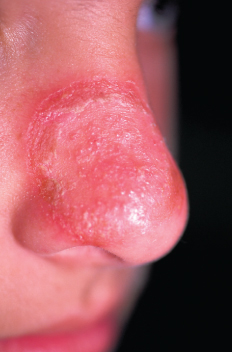
A 10-year-old boy presents with a 10-day history of a worsening rash on his nose. The patient says that the area is slightly itchy and a little tender. He denies rash elsewhere on his body.
What are you looking at here?
A. Impetigo.
B. Candidiasis.
C. Ringworm.
D. Contact dermatitis.
E. Acne.
Case 3: Dermatophyte infection
The sudden onset and dramatic appearance of the eruption could be misleading. A potassium hydroxide evaluation confirmed the diagnosis of a dermatophyte infection,C.
The lesions of impetigo and candidiasis would be expected to have a more golden crust. The typical clinical appearance of contact dermatitis and a history that implicated a causative agent were absent here. Acne of only 10 days’ duration does not resemble this lesion.
This patient’s ringworm responded quickly to a 1-week course of oral antifungal antibiotics in combination with a topical antifungal cream.
Case 4:
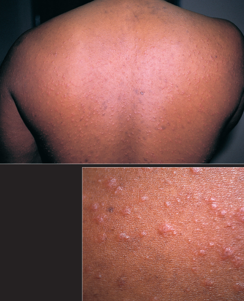
A 31-year-old man seeks evaluation of a pruritic papular eruption on his trunk of 2 weeks’ duration. He denies fever, malaise, and medication use.
Your first step is to . . .
A. Perform a skin biopsy.
B. Obtain a complete blood cell count and chemistry panel.
C. Perform a VDRL test.
D. Obtain a chest radiograph.
E. Obtain an antinuclear antibody titer.
Case 4: Pityriasis rosea
This is a typical presentation of pityriasis rosea, which usually manifests as oval-shaped, salmon-colored macules or erythematous papules. Since secondary syphilis can mimic pityriasis rosea, a VDRL test, C, can be useful; the other tests are of limited value. n