
Various Manifestations of Rheumatic Disorders
Photo Essay
Focus on Signs and Symptoms
 Case 1
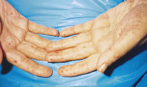
Rheumatoid Vasculitis
Ten weeks before presentation, this 55-year-old woman noticed decreased sensation in her feet and a bluish discoloration of her toes. These symptoms progressed rapidly, and pain and coldness in both feet increased in intensity. Her feet subsequently became gangrenous. Her seropositive arthritis had been diagnosed about 6 years earlier. The disease had been well controlled until about 10 weeks before this photograph was taken.
This image is a particularly good depiction of the livedo reticularis pattern involving the legs. The fishnet appearance of purple discoloration is frequently an indicator of vasculitis.
Rheumatoid vasculitis is a rare complication of rheumatoid disease and tends to affect patients with a long history of rheumatoid arthritis and a high rheumatoid factor titer. Most patients in this group would be expected to have rheumatoid nodules as well. Rheumatoid vasculitis may also cause hyperkeratotic papules around the elbows and knees, nail fold infarcts, deep cutaneous ulcers, and peripheral neuropathy or mononeuritis. ■
(Case and photograph courtesy of Dr David I. Wolf.)

Case 2
Idiopathic Dermatomyositis
A 76-year-old woman complained of progressive proximal muscle weakness; achy pain in the buttocks, thighs, and calves; and lilac discoloration of her eyelids, cheek, nose, knuckles, and fingernails.
Examination confirmed heliotrope skin changes and disclosed moderate weakness in the limb and girdle musculature. The patient’s levels of muscle enzymes, including creatine kinase, aldolase, aspartate aminotransferase, alanine aminotransferase, and lactate dehydrogenase, were elevated, as was her erythrocyte sedimentation rate. An electromyogram revealed increased exertional activity, fibrillation potentials, and a typical myopathic pattern. Muscle biopsy was performed, and the findings were consistent with inflammatory myositis. Evaluation for an underlying malignancy yielded negative results.
The diagnosis of idiopathic dermatomyositis was based on 4 criteria: clinical picture, electromyographic findings, elevated serum creatine kinase level, and biopsy report. The patient was treated with prednisone, 60 mg/d, and was given physical therapy. Three months later, her strength had improved by 80%. ■
(Case and photograph courtesy of Dr Gavin I. Awerbuch.)
Case 3
Progressive Systemic Sclerosis
For several months, a 70-year-old woman had had dysphagia, mild dyspnea on exertion, and the Raynaud phenomenon. Her skin was waxy and edematous; 2- to 10-mm pinkish spots had appeared on her fingers, palms, and oral mucous membrane over the past 2 weeks. These disappeared completely with pressure. Subcutaneous calcific deposits were present on the extensor surfaces of the forearms.
The spots are telangiectases of progressive systemic sclerosis. The Raynaud phenomenon is seen in about 90% of cases, and skin thickening, pigment changes, and internal organ fibrosis are important characteristics.
Patients with limited systemic scleroderma frequently have calcinosis cutis, the Raynaud phenomenon, esophageal dysfunction, sclerodactyly, and telangiectasia (CREST syndrome), as this woman did. In such patients (compared with those who have diffuse systemic scleroderma), skin tightening is confined to the hands and face; there is a lower risk of renal involvement, a higher risk of pulmonary hypertension, and an overall better prognosis. In diffuse systemic sclerosis, the course of visceral disease is more rapid, leading to death.
Laboratory findings disclose a variety of abnormalities. There may be mild anemia, hyper-gammaglobulin-emia (in 50% of patients), proteinuria, positive tests for antinuclear antibodies, presence of anticentromere antibody (in 50% of those with CREST syndrome and 1% of those with
diffuse systemic scleroderma), and Scl-70 antibodies (in 20% of those with CREST syndrome and 33% of those with diffuse disease).
Diseases that must be considered in the differential diagnosis include eosinophilic fasciitis (which is not associated with the Raynaud phenomenon, manifests peripheral blood eosinophilia, and responds to prednisone therapy); porphyria cutanea tarda; chronic graft-versus-host disease; and eosinophilia-myalgia syndrome secondary to tryptophan ingestion. ■
(Case and photograph courtesy of Dr Shanon D. Smith.)
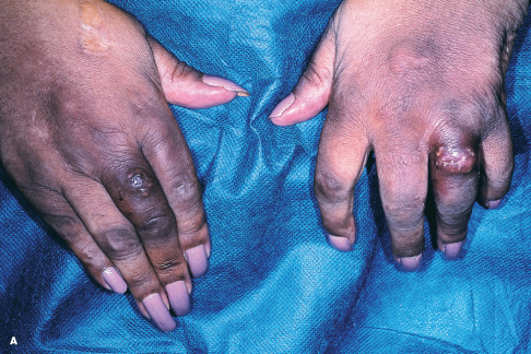
Case 4
Rheumatoid Nodules
A 65-year-old woman, who was confined to a wheelchair because of severe rheumatoid arthritis, was concerned about nodules that had erupted on her fingers and hands during the previous 3 weeks (A). Her medical history included colon cancer, chronic renal insufficiency, anemia, and hypertension. The nonpruritic nodules were painful when they began to form under the skin; however, once they erupted, the pain disappeared.
Four firm, irregular nodules at various stages of development were noted on the dorsa of the fingers and hands. One of the lesions had exuded a yellow-white chalky material from several locations. The patient reported the occurrence of similar nodules in the past; 8 months earlier, a lesion erupted over the left fifth metacarpophalangeal joint, and more recently, a nodule developed over the ulnar surface of the left forearm. Both lesions discharged moist, yellow material and resolved spontaneously within several days.
The patient’s medical history raised the possibility of cutaneous calcium deposition. However, roentgenograms of the hands revealed osteopenia and lytic lesions around the joints but no calcium deposits. Erosive and cystic changes were demonstrated at the proximal interphalangeal joints of the second through fifth fingers; similar changes were more prominent at the second and third metacarpophalangeal joint space of the right hand (top row) than of the left (bottom row). The diagnosis of rheumatoid nodules was confirmed clinically. Rheumatoid nodules are found in approximately one third of patients with rheumatoid arthritis. Usually, they are associated with more severe disease and a high rheumatoid factor titer. These lesions also occur in about 5% of persons with systemic lupus erythematosus. Most often located over bony prominences or extensor surfaces—notably on the forearms, elbows, knuckles, feet, and knees—the nodules tend to be deep and asymptomatic. The presentation of eruptive nodules in this patient was atypical. Intralesional corticosteroids were injected into the largest nodule, after which all of the developing and fully developed lesions disappeared completely. At the 4-month follow-up, no recurrence of lesions was noted despite the patient’s refractory arthritis. ■
(Case and photographs courtesy of Drs Jessica Krant and Yelva Lynfield.)