
Interactive Quiz: Persistent Cough in a Child

Welcome to Pulmonology Consultant's latest interactive diagnostic quiz. Over the next few pages, we'll present a case and ask you to make the diagnosis and treat the patient. Along the way, we'll provide details about the case, and at the end, we'll share the patient's outcome.
Ready to get started? >>
First, let’s meet the patient…
A 2-year-old boy presented to the emergency department (ED) with a persistent cough with production of green purulent sputum.
The patient’s medical history was significant for a febrile seizure and repair of mixed total anomalous pulmonary venous return with stenting of left and pulmonary vein confluences, as well as respiratory syncytial virus bronchiolitis 1 month before admission.
His primary care physician had prescribed 5 days of prednisolone out of concern for a cough that likely was a symptom of reactive airway disease. On the final day of corticosteroid therapy, the patient’s mother had noticed that the boy suddenly had developed difficulty breathing and felt feverish.
In the ED, the patient’s vital signs were as follows: temperature, 38°C; blood pressure, 98/57 mm Hg; heart rate, 147 beats/min; respiratory rate, 31 breaths/minute; and oxygen saturation, 94% on room air. Pertinent physical examination findings were abdominal breathing and mild subcostal retraction.
Are you correct? >>
Answer: Treat the patient with a bronchodilator
The patient was treated with a bronchodilator (albuterol-ipratropium combination) and 8 L/min of oxygen via a nasal cannula, in addition to 20 mL/kg normal saline bolus.
Findings of a subsequent physical examination included respiratory distress with tachypnea, subcostal and intercostal retractions with nasal flaring, and grunting, as well as symmetric coarse breath sounds. Cardiovascular examination findings were unremarkable.
Are you correct? >>
Answer: Chest radiograph
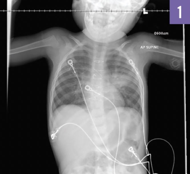
A chest radiograph (Figure 1) demonstrated a single well-circumscribed opacity within the left upper lobe.
Are you correct? >>
Answer: Round pneumonia
The patient received intravenous vancomycin and cefepime. Blood cultures eventually grew Streptococcus pneumoniae.
The following day, the patient was weaned from oxygen therapy and was stable on room air. The patient demonstrated excellent ability to tolerate an oral diet, and he was discharged to home to complete a total of 10 days of antibiotic (amoxicillin) therapy.
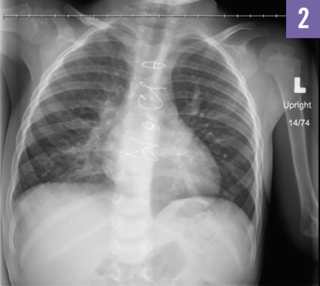
A follow-up chest radiograph after 4 months (Figure 2) was performed.
Are you correct? >>
Answer: No more treatment is necessary
The follow-up chest radiograph (Figure 2) showed resolution of pneumonia. Repeated radiographic studies in the pediatric population with round pneumonia after appropriate antibiotic therapy have been shown to be of little value in patients whose clinical symptoms are responding well to treatment.
Are you correct? >>
Answer: No
Round pneumonia is a common clinical entity in children and adolescents. Studies show that the mean age at diagnosis ranges from 3.3 to 5 years, with a nearly equal sex ratio. When combined with the correct clinical picture of pneumonia, it is a relatively benign radiologic finding.
To read the full case report, see:
Wetzig A, Barnes K, Gourishankar A. Round pneumonia. Consultant. 2018;58(11):320-321. https://www.consultant360.com/article/pulmonology/pneumonia/round-pneumonia.