
Pityriasis Rubra Pilaris
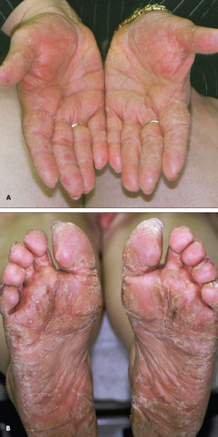 This long-standing eruption on a 52-year-old woman’s palms (A) and soles (B) had resisted treatment with oral and topical antifungals, corticosteroid creams, and antibiotics. Her symptoms included pruritus and pain from the fissures that developed at the involved sites. The eruption had been present for about 10 years. Her father had a similar condition; however, the diagnosis was unknown.
A marked hyperkeratotic effect was noted on the palms and soles, along with a yellowish color. The nails, scalp, elbows, knees, and nails were uninvolved. Biopsy showed changes consistent with pityriasis rubra pilaris (PRP).
PRP is considered to be part of the papulosquamous family of disorders, which also includes psoriasis. The diagnosis is usually clinical, since histopathologic findings are not pathognomonic. The adult type is of acute onset and involves cephalad to caudad spread of lesions; ectropions; follicular papules on the dorsal aspects of fingers, and extensor aspects of wrists and thighs; and hyperkeratotic palms and soles with an orange hue. Often, there is progressive erythroderma with islands of sparing. This patient had an atypical, familial form of PRP.
Treatment is with oral retinoids, most commonly isotretinoin. Because PRP is notoriously difficult to treat, many other treatments have been tried, including antibiotics, methotrexate, phototherapy, and cyclosporine.