
Do You Know Your Nevi?
Case 1:
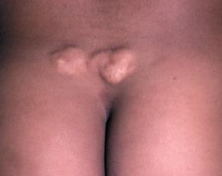
The parents of a 3-year-old girl seek medical evaluation of the nodules on their daughter’s back. The lesions have been present since birth and have grown with the child.
What is your clinical impression?
(Answer on next page.)
Case 1: Nevus lipomatosus
This is nevus lipomatosus; the group of soft yellow nodules have coalesced to form a plaque over the patient’s sacrum. These nevi have a predilection for the lower back and the upper thighs. This diagnosis is made clinically and, if necessary, can be confirmed by a biopsy.
Nevus lipomatosus results from the abnormal presence of adipose tissue in the dermis. Typically, these benign tumorlike lesions are noted at birth or in childhood; over time, they can grow and may become pedunculated. This nevus can be removed by simple excision.
(Case and photograph courtesy of Dr Caron Grin.)
 Case 2:
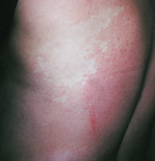
A pigmented lesion has been present on a teenager’s back for a year, but the surrounding circle of depigmentation developed recently after a sunburn. The lesion and surrounding skin are neither pruritic nor painful.
Is biopsy warranted?
(Answer on next page.)
Case 2: Halo nevus
This is a halo, or Sutton, nevus. These lesions usually arise during adolescence; multiple halo nevi are often present.
Initially, a halo of depigmentation appears around an existing nevus. The central nevus often disappears over time, and an area of depigmentation remains. This area may eventually repigment.
Histologic examination of a halo nevus reveals a striking lymphocytic infiltrate admixed with nevus cells in the dermis and at the dermoepidermal junction, with a loss of epidermal melanocytes in the depigmented halo area.
 Frequently, these nevi are first noted during the summer because the halo—unlike adjacent normal skin—does not tan. These lesions are usually benign when seen in adolescents; however, use the “ABCD” lesion assessment criteria (Table) to determine whether a biopsy is warranted. In an adult, the development of an isolated pigmented lesion surrounded by a halo can be benign, but excision is recommended to rule out the remote possibility of a halo melanoma.
REFERENCE:
1. Friedman RJ, Rigel DS. The clinical features of malignant melanoma. Dermatol Clin. 1985;3:
271-283.
(Case and photograph courtesy of
Joe Monroe, PA-C.)
 Case 3:
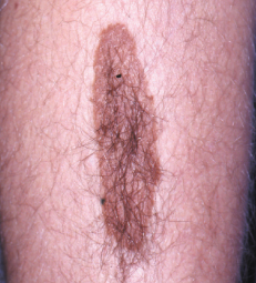
A 2.5 3 8-cm hairy, pigmented lesion is noted on a 15-year-old boy’s lower leg; the lesion has been present since birth. The size of the birthmark has not increased disproportionately over time.
Is it likely that this lesion is malignant?
(Answer on next page.)
Case 3: Congenital hairy nevus
The patient has a congenital hairy nevus. Congenital nevi are present at birth and can vary in size from a few millimeters to many centimeters. The lesions are usually brown or black but may be red or pink. They are often flat or barely raised at birth and thicken with age. The surface of the nevus may become nodular; the hair that can arise is usually coarse.
Giant congenital nevi are defined as larger than 20 cm. One that covers a large portion of the trunk is referred to as a bathing trunk nevus.
The risk of melanoma in small congenital nevi is controversial; however, most experts consider it to be relatively low. Because giant hairy congenital nevi pose a higher risk of melanoma, referral to a surgeon for consideration of removal is warranted. Small congenital nevi may be followed by close observation, or they may be excised to eliminate any risk of malignant transformation.
FOR MORE INFORMATION:
Weedon D. Skin Pathology. London: Churchill Livingstone; 2002:815-816.
(Case and photograph courtesy of Dr Robert P. Blereau.)
 Case 4:
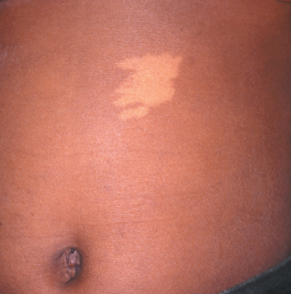
An irregular pale white macule has been present on the trunk of an 8-year-old boy since birth. The area is asymptomatic. The patient has no other similar lesions.
What do you suspect—and how will you narrow the differential?
(Answer on next page.)
Case 4: Nevus depigmentosus
Nevus depigmentosus is a birthmark that occurs in approximately 1 in 125 persons. It can present as a single, well-demarcated hypomelanotic macule that ranges from 0.5 to 10 cm. Less often, the distribution is segmental or systematized (multiple streaks following the lines of Blaschko).
A nevus depigmentosus can be distinguished from a nevus anemicus by diascopy, which is performed by pressing a clear glass slide on the lesion. The boundary of the nevus depigmentosus remains visible during this procedure, whereas the border of the nevus anemicus disappears.
The differential diagnosis includes the ash-leaf spot of tuberous sclerosis. When a child presents with multiple lesions and/or additional signs or symptoms that suggest tuberous sclerosis, such as seizures or facial angiofibromas, further evaluation, including diagnostic imaging, is warranted. The presence of a single lesion in an asymptomatic child requires observation only.
Because this patient’s nevus was an isolated lesion, follow-up and reassurance were sufficient.
FOR MORE INFORMATION:
Ackerman AB, Kerl H, Sanchez J. A Clinical Atlas of 101 Common Skin
Diseases: With Histopathologic Correlation. New York: Ardor Scribendi; 2000.
Dover JS, Jackson BA. Pocket Guide to Cutaneous Medicine and Surgery.
Philadelphia: WB Saunders Co; 1996.
(Case and photograph courtesy of Dr Charles E. Crutchfield III.)
 Case 5:
A 32-year-old man presents with a hypopigmented lesion on his left flank. The lesion has been present since infancy.
What steps will you take to
determine the cause of the area of depigmentation?
(Answer on next page.)
Case 5: Nevus anemicus
When pressed with a glass slide, a nevus anemicus becomes indistinguishable from the surrounding normal skin. Friction or applications of heat or cold to the nevus do not produce erythema within the lesion; however, the surrounding skin does respond. The absence of erythema is attributed to blood vessel constriction that is secondary to increased blood vessel sensitivity to catecholamines.
A Wood lamp examination accentuates patches of vitiligo, which is in the differential; whereas a nevus anemicus is invisible under the lamp. No treatment is necessary for this nevus.
(Case and photograph courtesy of Dr Raymond Kuwahara.
 Case 6:
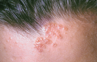
An 8-year-old girl is brought to the office for evaluation of a lesion on the back of her neck, which was first noted a few years earlier. Examination reveals multiple comedones grouped together.
Can you identify this lesion?
(Answer on next page.)
Case 6: Nevus comedonicus
A nevus comedonicus is a patch-like collection of flesh-colored papules, each with a comedo-like dilated center. It may be congenital or acquired. The face, neck, upper arm, and chest are sites of predilection. These lesions are usually of cosmetic concern only.
Because of the typical comedonal lesions, these nevi are generally relatively easy to diagnose. The differential includes nevus sebaceus, molluscum, and acne; if the diagnosis is in doubt, a punch biopsy may be performed.
Removal for cosmesis can be accomplished by excision or carbon dioxide laser ablation.
(Case and photograph courtesy of Joe Monroe, PA-C.)
 Case 7:
For as long as he can remember, a 40-year-old man has had a red-purple discoloration on the right side of his face, head, and neck. He has never had any symptoms associated with this nevus.
What are your thoughts about this lesion?
(Answer on next page.)
Case 7: Nevus flammeus
A port-wine stain (or nevus flammeus) is a congenital vascular malformation of mature dilated capillaries in the entire dermis; the condition is not genetically transmitted. Most often, these lesions occur on the face but may appear on any part of the body. They need to be differentiated from benign flame nevi, which commonly occur in newborns and are known as “salmon patches,” “stork bites,” and “angel kisses.” Port-wine stains are flat and smooth at birth and—unlike benign flame nevi, which fade spontaneously over a few years—tend to become papular, nodular, and darker with age.
A port-wine stain can occur as an isolated finding or as part of a syndrome. When a stain involves both sides of the face or both the upper and lower eyelids (the first branch of the trigeminal nerve), there is a risk of vascular malformations of the ipsilateral leptomeninges with associated mental retardation, glaucoma, and seizures (Sturge-Weber syndrome) and congenital cataracts.
The disfigurement caused by port-wine nevi is often psychologically devastating. Makeup applied to affected areas may significantly improve a patient’s appearance and self-esteem. Tunable dye laser therapy performed in early childhood is the most beneficial treatment.
(Case and photograph courtesy of Dr Robert P. Blereau.)