
A Mouthful of Oral Lesions, Part 2
Mouthful of Oral Lesions (Part 1)
Case 1:
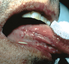
Several punch biopsies of this tongue lesion were done to rule out malignancy.
What does it look like to you?
Answer on next page
Case 1: Lichen planus
This is ulcerated lichen planus of the tongue in a 60-year-old man. The ulcerations eventually responded to topical and intralesionally injected corticosteroids.
(Case and photograph courtesy of Dr Ernst Epstein.)
Continued on next page
Case 2:
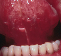
These sublingual lesions developed in a 25-year-old man.
What might be the cause?
Answer on next page
Case 2: Sublingual condylomata
These are sublingual condylomata. They were excised with scissors and the bases of the lesions were electrodessicated; they did not recur. The patient also had multiple penile and perianal condylomata, which were successfully removed.
(Case and photograph courtesy of Dr Marvin I. Lepaw.)
Continued on next page

Case 3:
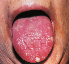
This lobulated lesion developed in a 42-year-old woman more than a year before this photograph was taken.
Although it bled occasionally from minor trauma, there were no other symptoms.
What does this lesion suggest, and what does it portend?
Answer on next page
Case 3: Pyogenic granuloma
This slow-growing, warty-surfaced lesion proved to be a pyogenic granuloma, which commonly involves the gingivae, lips, and buccal mucosa. This woman had no other granulomatous lesions, however.
The granuloma was removed by radical surgical excision. It did not recur.
(Case and photograph courtesy of Dr Frederick A. Saverice.)
Continued on next page
Case 4:
A 75-year-old woman was hospitalized with dyspnea on exertion and fatigue. Glossitis and pallor were observed.
To what diagnosis do these clinical findings point?
Answer on next page
Case 4: Pernicious anemia
Glossitis and pallor suggested pernicious anemia. The hematocrit was 25%; mean corpuscular volume (MCV), 125 µm3; white blood cell (WBC) count, 1200/µL; platelet count, 120,000/µL; reticulocytes, 27,500/µL; vitamin B12, 64 pg/mL; folic acid, 8.2 ng/mL; total bilirubin, 1 mg/dL; and lactic dehydrogenase, 639 IU/L (normal values, 100 to 250 IU/L). Macro-ovalocytosis and anisocytosis with neutrophil hypersegmentation were seen in the blood smear; giant promyelocytes were found in the bone marrow aspiration. These findings confirmed the diagnosis of megaloblastic anemia.
Parenteral therapy with vitamin B12 was initiated. Six days later, the number of reticulocytes had increased markedly to 100,000/µL; hematocrit was 36%; MCV, 100 µm3; WBC count, 3800/µL; and platelet count, 145,000/µL. The patient was released from the hospital; oral vitamin B12 therapy was continued at home.
(Case and photograph courtesy of Drs Evangelos Rizos, George Alexadridis, and Moses Elisaf.)