
Man With Epigastric Pain and Melena
A 67-year-old man is admitted to the hospital after he experienced 4 or 5 episodes of melena in the past 24 hours. He has had no emesis, but complains of moderate epigastric pain.
HISTORY
He has had similar though less severe epigastric pain in the weeks prior to this episode. He had a similar event, but with less passage of melena, 4 years earlier. At that time, he underwent endoscopy, and he recalls being told that he had “ulcers but no tumor.”
Since then he has taken proton pump inhibitors intermittently, particularly when he experiences epigastric discomfort. In addition, he takes an angiotensin-converting enzyme inhibitor and a diuretic daily for long-term essential hypertension. He smokes cigars occasionally and does not drink alcohol.
PHYSICAL EXAMINATION
The patient appears pale. He is afebrile with a supine pulse of 104 beats per minute (bpm) and blood pressure (BP) of 95/70 mm Hg, which changes to 120 bpm and 80/60 mm Hg when he is upright. There is no scleral icterus, but the mucosae are pale. The chest is clear, and the heart examination reveals tachycardia and a summation gallop.
Bowel sounds are audible. Moderate epigastric tenderness is noted on deep palpation, but there is no rebound. No hepatosplenomegaly is detected. The rectal examination results are normal, but grossly melanotic stool, which is strongly positive for occult blood, is present. The remainder of the physical examination is normal.
LABORATORY AND IMAGING RESULTS
White blood cell count is 14,300/µL; platelet count is normal. Hemoglobin level is 8.9 g/dL with a normal mean corpuscular volume. Liver function test results and prothrombin time are normal. Creatinine level is 1.0 mg/dL, and blood urea nitrogen (BUN) level is 28 mg/dL. An ECG shows sinus tachycardia and left ventricular hypertrophy by voltage criteria with non-specific ST-T changes in leads I, aVL, and V4 through V6.
Which of the following is the most accurate statement about the management of this patient’s condition?
A. Regardless of endoscopic findings, the patient has a significant risk of mortality.
B. He should receive intravenous erythromycin as a promotility agent to improve endoscopic visualization.
C. A nasogastric tube with lavage is needed to most accurately stratify risk.
D. Endoscopy and endoscopic therapy should be delayed 72 hours.
Correct Answer: A
This patient exhibits manifestations typical of bleeding peptic ulcer, a common and dramatic condition that accounts for roughly half of all episodes of upper GI hemorrhage. This should be the presumed diagnosis here.
PROGNOSTIC FEATURES IN PEPTIC ULCER DISEASE
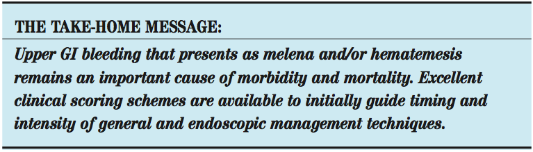
The prognosis overall for patients with bleeding peptic ulcer is quite good. Yet, despite all of the gains accrued in the knowledge of pathophysiology (ie, Helicobacter pylori), diagnostics (endoscopy), and therapeutics (proton pump inhibitors and endoscopy), the overall mortality remains little changed at 5% to 10%.1,2 This patient’s GI bleeding manifested as melena, which occurs in about 20% of cases. Hematemesis is the presentation in 30% of cases, while 50% exhibit both.1
The history of recurrence is typical and had been the rule prior to the advent of effective therapy to eradicate H pylori. Despite the impressive advances in endoscopy procedures for diagnosis, prognosis, and even therapy, many important prognostic features to determine poor peptic ulcer disease outcomes (ie, need for urgent endoscopy, rebleeding, and death) remain critical. This patient demonstrated several of these features.
Two important and frequently used clinical risk stratification tools for upper GI hemorrhage are the Blatchford and Rockall scores.2 This patient manifested several clinical clues consistent with at least moderate risk of rebleeding or death. His age (older than 60 years), systolic BP (less than 100 mm Hg), hemoglobin level (less than 10 g/dL), pulse (more than 100 bpm), BUN (higher than 24 mg/dL), and melena accrue 14/23 Blatchford and 3/7 Rockall points prospectively using these scoring systems.
He is 67 years old, and poor outcomes are very significantly concentrated in patients older than 60 years. In a large older study of more than 700 patients with bleeding ulcers, the mortality in patients under 60 was 0.5%, while in those older than 60 it was 10%, a 20-fold difference.3 Similar findings appeared in a more recent study as well.4 Thus, choice A is correct. Clinical criteria at triage establish that he is at significant risk for rebleeding and death.
DIAGNOSTIC AND THERAPEUTIC MANEUVERS
Because of his clinical parameters, trials and consensus have shown that early endoscopy (within 24 hours of presentation) is recommended.5 This allows for prompt identification of high-risk lesions, such as spurting blood, adherent clots, and visible vessels, which all increase rebleeding and death and thus guide management regarding the use of endoscopic hemostasis, intensive care placement, and timing of discharge. Thus, choice D is too conservative and is not appropriate here.
Conversely, the time-honored maneuver of nasogastric tube placement with lavage (choice C), though useful diagnostically to confirm that hemorrhage is likely proximal to the ligament of Treitz, is currently thought to provide little other incremental information and does not stop bleeding or prevent recurrences.2 Thus, choice C also is incorrect.
Another interesting maneuver, using promotility agents such as intravenous erythromycin to improve visualization (which it indeed does), has not been shown to improve either endoscopic diagnostic yield or overall outcome.2 Again by meta-analysis and consensus, such agents should not be used routinely,5 and choice B is not correct.
OUTCOME OF THIS CASE
The patient was resuscitated with fluids and was transfused with packed red blood cells. His vital signs stabilized as did his hemoglobin in the 9.5 to 10.5 g/dL range. He underwent endoscopy within 24 hours of presentation. During endoscopy, a 1.5-cm ulcer on the lesser curvature, associated with adherent clot, was identified and treated with thermal coagulation. Biopsy was negative for tumor, and H pylori testing and staining results are pending. He spent 24 hours in ICU observation and was discharged on day 4 on a proton pump inhibitor regimen.
1. Laine L, Peterson W. Bleeding peptic ulcer. N Engl J Med. 1994;331:717-727.
2. Gralnek IM, Barkun AN, Bardou M. Management of acute bleeding from a peptic ulcer. N Engl J Med. 2008;359:928-936.
3. Branick FJ, Coleman SY, Fok PJ, et al. Bleeding peptic ulcer: a prospective evaluation of risk factors for rebleeding and mortality. World J Surg. 1990;14:262-270.
4. Khuroo MS, Yattoo GN, Javid G, et al. A comparison of omeprazole and placebo for bleeding peptic ulcer. N Engl J Med. 1997;336:1054-1058.
5. Barkun AN, Bardou M, Kulpers EJ, et al. International consensus recommendations on the management of patients with nonvariceal upper gastrointestinal bleeding.Ann Intern Med. 2010;152:101-113.