
Liver Abscess Related to Foreign Body
Authors:
Gentiana Bakaj, MD
East Carolina University/Pitt County
Memorial Hospital, Greenville, NC
Mahfuzul Haque, MD, FRACP
Duke University
Citation:
Bakaj G, Haque M. Liver abscess related to foreign body. Consultant. 2013;53(3):195.
An 82-year-old African American woman presented to the emergency department with abdominal pain of 5 days’ duration; the pain was aggravated by movement and relieved somewhat by sitting down. She denied dysphagia, odynophagia, nausea, vomiting, or change in bowel movement.
The patient’s temperature was 39.2°C (102.6°F). Other significant physical findings were a heart rate ranging from 102 to 109 beats per minutes, blood pressure ranging from 116/53 to 151/60 mm Hg, bilateral expiratory wheezing, and hypoactive bowel sounds.
Laboratory studies revealed a white blood cell count of 13,900/µL with 85% neutrophils and less than 10% bands; hematocrit, 31.4%; total bilirubin, 1.4 mg/dL; and aspartate aminotransferase, 45 U/L. A CT scan of the abdomen (A) showed a 7.2 3 5.6 cm complex, septated, low attenuated mass with internal gas compatible with an abscess in the left lobe of the liver adjacent to the gastric wall. A linear, calcified density was noted; it extended from the collection through the wall of the gastric antrum, suggesting a foreign body. The patient underwent esophagogastroduodenoscopy with removal of a 3.7-cm fish bone (B) and percutaneous drainage of the abscess under ultrasound and fluoroscopic guidance.
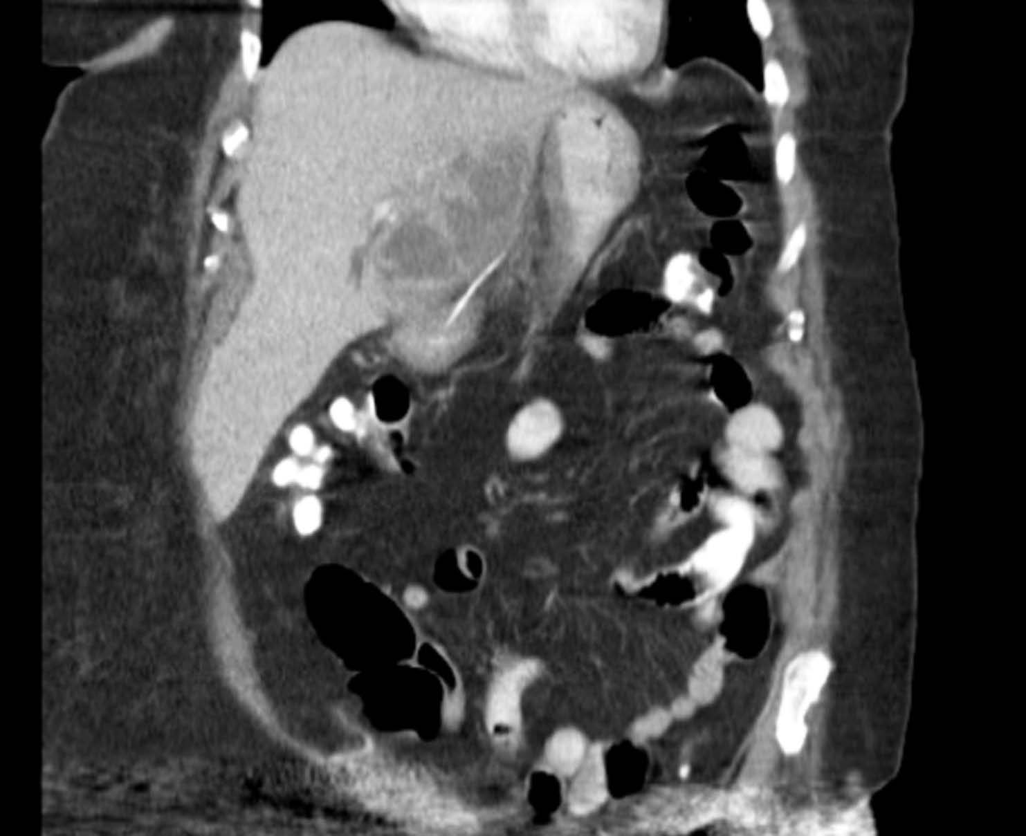
Figure A. Computed tomography scan of the abdomen.
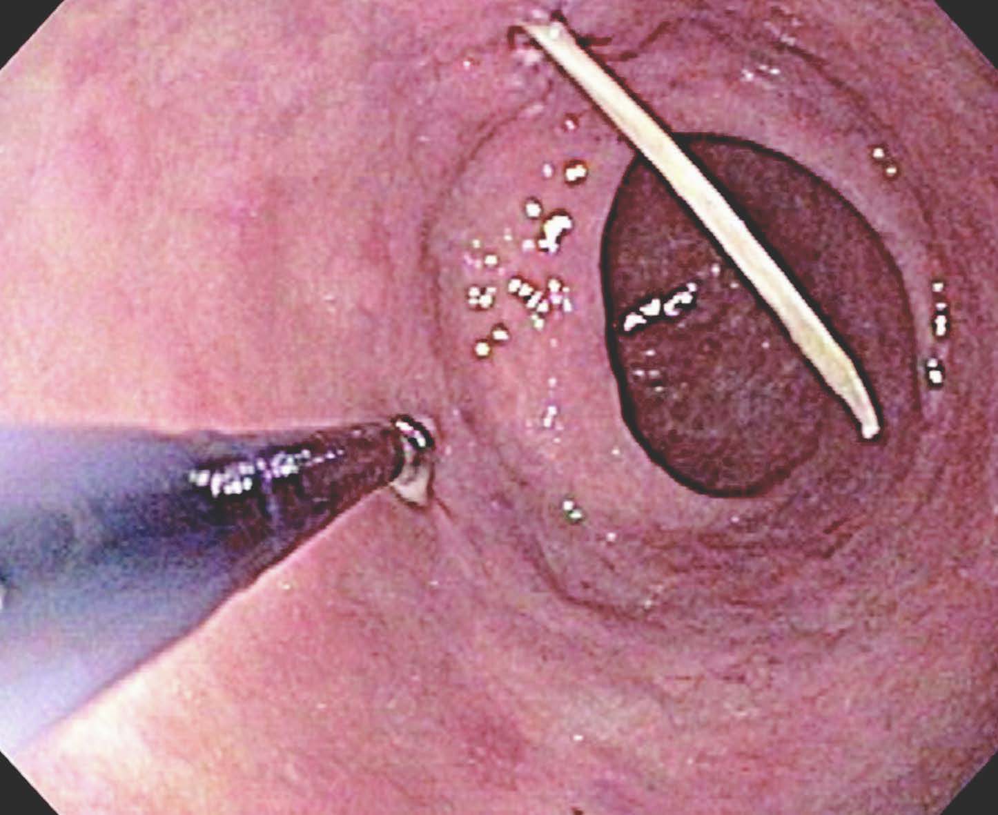
Figure B. Esophagogastroduodenoscopy showing fishbone impaction.
A 10 French all-purpose drainage catheter was placed within the cavity, and 100 mL of purulent, foul-smelling material was obtained and sent for analysis. A J tube was placed, which drained about 35 mL of serous discharge daily. Gram staining showed many gram-positive cocci and rare gram-negative rods. Culture grew 41 viridans streptococci. Blood culture remained negative.
During the hospital stay, the patient received doripenem and was discharged home on ceftriaxone, 1 g daily for 14 days. A repeat CT scan after drainage showed a 3.7 × 3.0 × 3.3 cm low attenuation lesion in the lateral segment of the left lobe consistent with inflammatory changes and minimal residual fluid. The drain was removed after 16 days, and the patient remained asymptomatic.
FOR MORE INFORMATION:
- Aygencel G, Dizbay M. Severe systemic sepsis due to pyogenic liver abscess. Ir J Med Sci. 2012;181(2):289-291. doi: 10.1007/s11845-010-0611-2.2010 Oct 17.
- Gao HN, Yuan WX, Yang MF, et al. Clinical significance of C-reactive protein values in antibiotic treatment for pyogenic liver abscess. World J Gastroenterol. 2010;16(38):4871-4875.