
Leisure Time Lesions

Seabather’s Eruption
This 10-year-old boy presented for evaluation of a rash that developed during a spring vacation on Florida’s Atlantic coast. After he had been swimming in the ocean, a pruritic, erythematous, papular rash developed on his trunk, axillae, and groin. Approximately 24 hours after the onset of the rash, he experienced malaise, chills, and a sore throat. His past medical history was unremarkable. He had been fully immunized and had had varicella infection.
This boy has seabather’s eruption, a pruritic dermatitis that occurs after exposure to seawater that contains certain species of jellyfish—particularly the larval form of the thimble jellyfish (Linuche unguiculata). The larvae are found in seawater in areas where this eruption is endemic, such as the Caribbean and the Atlantic coast of Florida, where this boy had vacationed. The jellyfish are found in these warm waters from March through August, with peak concentrations in May and June. The first written reports of this disease came from Florida’s Delray Beach.
The jellyfish larvae contain venom-filled barbs that fire their venom into the skin when the larvae become trapped under a bathing suit. Changes in osmotic pressure, such as air-drying or showering with fresh water, also trigger the barb-firing process. The venom causes a local toxic reaction and can precipitate a systemic hypersensitivity reaction. The local reaction—an erythematous, pruritic, papular dermatitis—may be
especially heavy on the skin under the bathing suit and in areas covered with hair, such as the axillae. It appears 4 to 48 hours after exposure to infested seawater. Systemic symptoms are more common in children than adults and include malaise, fever, sore throat, abdominal pain, headache, cough, and diarrhea.
Both the rash and systemic symptoms generally resolve in 1 to 2 weeks. Treatment is supportive and includes antihistamines and topical corticosteroids. Systemic corticosteroids are reserved for severe cases. The best preventive measure is to avoid swimming in infested water altogether. Those who choose to swim in potentially infested waters need to be aware that the wearing of T-shirts for ultraviolet protection increases the risk of getting seabather’s eruption. Bathers should instead use a waterproof sunblock for ultraviolet protection. While sunblock affords no protection against the jellyfish larvae, it does not increase the risk of trapping the larvae against the skin, as T-shirts do. Bathers should shower as soon as possible after swimming, without their bathing suits, which should be washed in detergent and heat-dried.
(Case and photograph courtesy of Dr Mary L. Sy and Dr Gary P. Williams.)

Hot Tub Folliculitis
This 28-year-old woman presented with pruritic, erythematous macules that had developed around the hair follicles on her legs and buttocks 4 days after she used a hot tub with friends. Each lesion had a central vesicle or pustule.
Hot tub folliculitis can develop from 6 hours to 8 days after exposure in a communal hot tub. The offending organism is Pseudomonas aeruginosa, which can cause an eruption on any part of the body exposed to water. Although the rash is usually itchy, other symptoms or complications are rare. Treatment is not necessary, since the rash usually resolves spontaneously; this patient’s rash disappeared within 10 days or so. Folliculitis can also result from the use of a wet suit.
(Case and photograph courtesy of Dr David I. Wolf.)
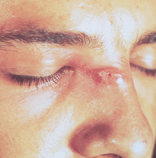
Swimming Pool Granuloma
This lesion developed 6 weeks after the patient had scraped his nose while swimming in a pool. Culture grew Mycobacterium marinum, an acid-fast organism that can be found in some home aquariums as well as in contaminated swimming pools.
(Case and photograph courtesy of Dr Carmen C. Thomas.)
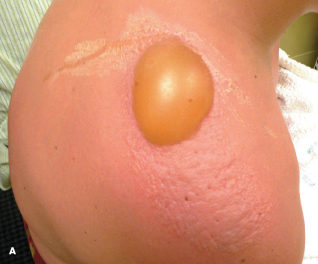 Second-Degree Sunburn
After spending a day at the beach, a 12-year-old boy presented with a second-degree sunburn and blisters on his shoulders (A). According to his mother, he had used sunscreen. The child was not taking any medications. Except for a significant number of moles, his skin was normal.
Because the majority of one’s lifetime exposure to sunlight occurs by age 18 years, and because sunburn at any age increases the risk of melanoma, sun protection must be emphasized to patients and their parents. Advise parents to begin protecting their children from sun exposure early, preferably in infancy, and to continue year-round. The use of sunscreen with a sun protection factor (SPF) of at least 15 to 30, depending on skin type, that protects against both UV-A (responsible for phototoxic and photosensitive drug reactions) and UV-B is recommended.
Advise parents to:
•Apply a liberal amount of sunscreen 30 minutes before sun exposure.
•Reapply sunscreen every 2 hours.
•Use waterproof sunscreen when the child will be in the water, exercising, or sweating.
 In 1999, the American Academy of Pediatrics recommended that for infants younger than 6 months, sunscreen may be applied to areas such as the face and dorsa of hands that may not be adequately covered by clothing.
The incidence of skin cancer—the most common, curable, and preventable form of cancer—is increasing rapidly because of increased longevity and exposure to UV light. Other risk factors for skin cancer include:
•Fair skin and light eyes, red or blonde hair.
•Tendency to burn easily and tan with difficulty.
•Tanning. (This is unhealthful and can induce cancer as well as cause aging and wrinkling; there is no such thing as a healthy tan.)
•Many moles (50 or more) and freckles.
•A history of sunburn (2 or 3 times in childhood).
•Family history of cancer.
•Certain genetic disorders, such as xeroderma pigmentosa and erythropoietic protoporphyria.
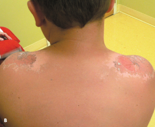
A typical sunburn—erythema that starts 2 to 6 hours after exposure to UV-B and peaks between 12 and 36 hours—resolves within 5 days. Cool compresses, refrigerated topical aloe, and hydrocortisone were applied to this patient’s shoulders and back, and he was given ibuprofen for the pain. After a week, the sunburned area had begun to heal (B).
(Case and photographs courtesy of Dr Bhagwan Das Bang.)

Swimmer’s Itch
A 6-year-old girl (A) and an 11-year-old boy (B) each presented for evaluation of an erythematous, pruritic, papular rash that developed after swimming in a
Wisconsin lake. Each child was otherwise completely healthy.
Both children have cercarial dermatitis, commonly known as swimmer’s itch. This is a hypersensitivity reaction that occurs after exposure to schistosome
larvae in freshwater lakes. Schistosome eggs (of trematodes, genera Schistosoma and Trichobilharzia) are passed into freshwater lakes via bird feces. The eggs hatch and develop into the larval stage called miracidia, which infect their intermediate host, freshwater mollusks. The miracidia transform into a second larval form called cercaria, which leave the mollusks and infect birds either by being ingested or penetrating their skin. Cercaria can also penetrate human skin and may infect persons swimming in infested water. The cercaria are unable to complete their life cycle in humans and die in the epidermis.
Although most common in the Great Lakes region of the United States, outbreaks occur during the summer months throughout the world. As the skin dries and the cercaria penetrate the epidermis, patients often describe the initial itching sensation. Small macules may be visible transiently at the site of penetration. Several days to 2 weeks after initial exposure, a mild to moderately pruritic, erythematous, papular, sometimes pustular eruption develops on any skin that had been exposed to infested water. In sensitized persons, the eruption develops within hours of exposure. The onset and intensity of the eruption correspond to the degree of sensitization. The rash peaks within 48 to 72 hours and usually resolves within 1 week.
Treatment is supportive and consists of oral and topical antihistamines and topical corticosteroids. Anthelmintic therapy is not indicated for this self-limited condition. Preventive measures include avoiding exposure to fresh water that contains mollusks and toweling off immediately after swimming (since penetration into the skin occurs during evaporation).
(Case and photographs courtesy of Dr Mary L. Sy and Dr Gary P. Williams.)

Cutaneous Larva Migrans
A linear, severely pruritic rash erupted on the lateral and plantar aspects of the left foot of a 72-year-old woman who had recently been camping on a Caribbean beach. Antihistamines and locally applied antifungal agents
offered no relief.
A biopsy of the lesion was unrevealing. A complete blood cell count showed eosinophilia of 12%. The travel history and clinical presentation of a wavy, meandering track on the foot confirmed the diagnosis of cutaneous larva migrans, or creeping eruption. The patient had been infected with the larva of Ancylostoma braziliense as she walked barefoot on the Caribbean beach. The ova-contaminated feces of dogs and cats are deposited on the sand, where the eggs hatch; the larvae may then penetrate the basal area of human skin.
This patient was given oral ivermectin. The rash and symptoms cleared completely.
(Case and photograph courtesy of Dr Navin M. Amin.)
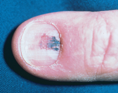 Sports-Related Nail Hemorrhage
This man’s thumb was struck by a racquetball, proximal to the cuticle, 12 weeks before the photograph was taken. He experienced only slight pain at the time of
injury. Within 12 hours, a bruise appeared in the soft tissue, without visible change in the nail. Discoloration persisted, and emerging nail showed hemorrhage
beneath the nail plate. The hemorrhage progressed distally with nail growth until normal nail began to grow beyond the cuticle.
Such hemorrhage can closely resemble melanin. Melanonychia may be caused by such diseases as lichen planus, as well as by drug reactions, AIDS, and other conditions, but more than one nail is usually affected. It is crucial to exclude subungual melanoma, and biopsy must be done unless that diagnosis can be eliminated unequivocally. Fortunately, the history of trauma followed by bruising usually establishes the diagnosis, although this is not always the case. For example, subungual hemorrhage may occur asymptomatically beneath a great toenail in joggers, even with properly fitted shoes; it is even more likely in persons wearing shoes that are too short.
(Case and photograph courtesy of Dr Wilfred E. Wooldridge.)