
Peer Reviewed
A Woman With a Warty Wrist
Authors:
Andrew Goodbred, MD
Associate Program Director, St. Luke’s Family Medicine Residency–Anderson, St Luke’s University Hospital Network, Easton, Pennsylvania
Satinderpal Kaur, MD
Resident, St. Luke’s Family Medicine Residency, St Luke’s University Hospital Network, Bethlehem, Pennsylvania
Citation:
Goodbred A, Kaur S. A woman with a warty wrist [published online December 2, 2019]. Consultant360.
A 47-year-old woman with no significant medical history presented with a mass over the volar aspect of her left wrist. The patient said that the mass had started growing 3 years ago, with a rapid increase in size over the past 6 months. She reported sharp pain and a pinching sensation at the site of the mass. She reported having occasional bleeding, and she had difficulty with wrist flexion and extension but was able to move her fingers. She denied numbness and tingling in her hand. She denied fever, weight loss, trauma, and exposure to any new chemical. The remainder of findings of a review of systems were unremarkable.
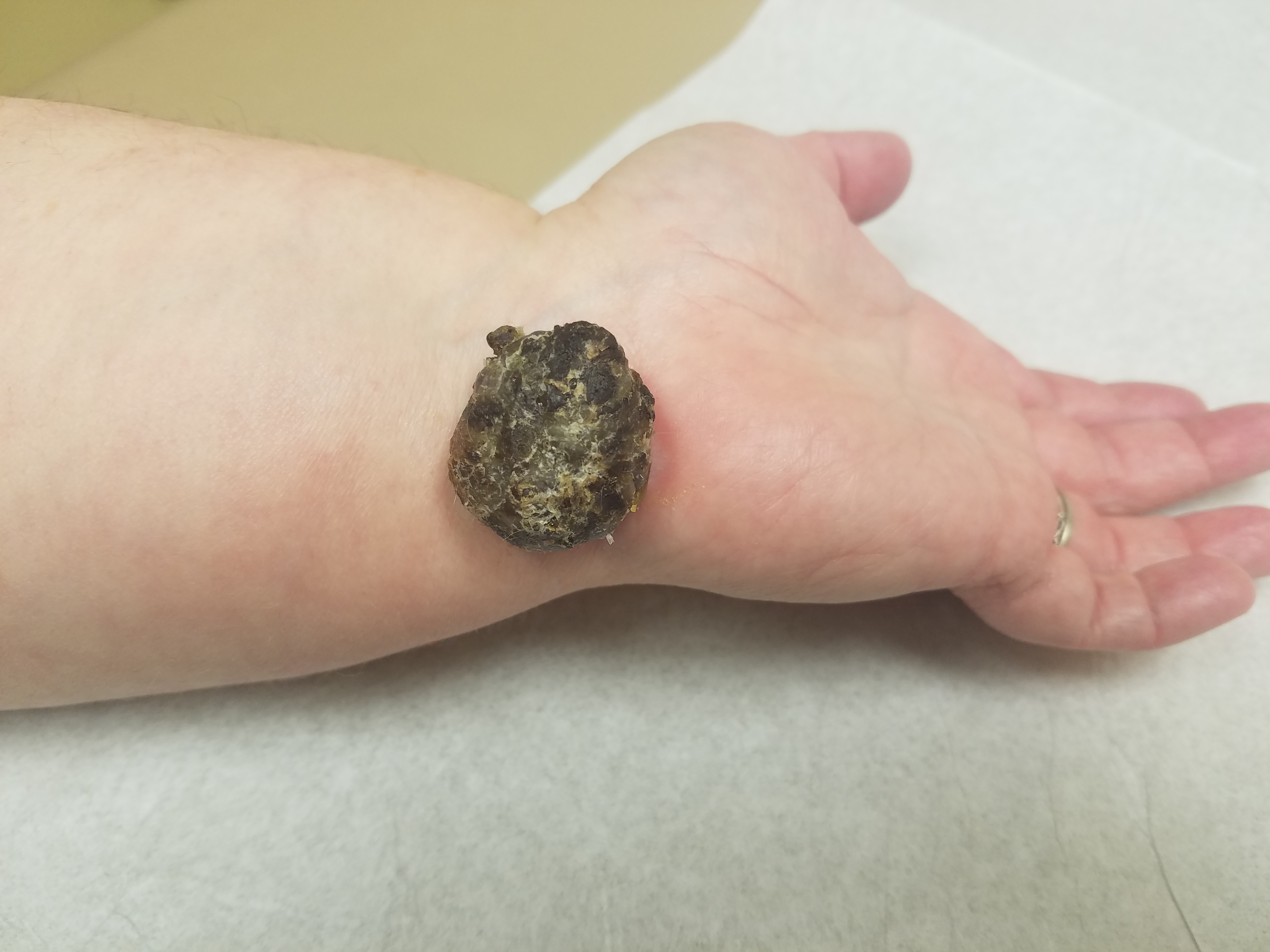
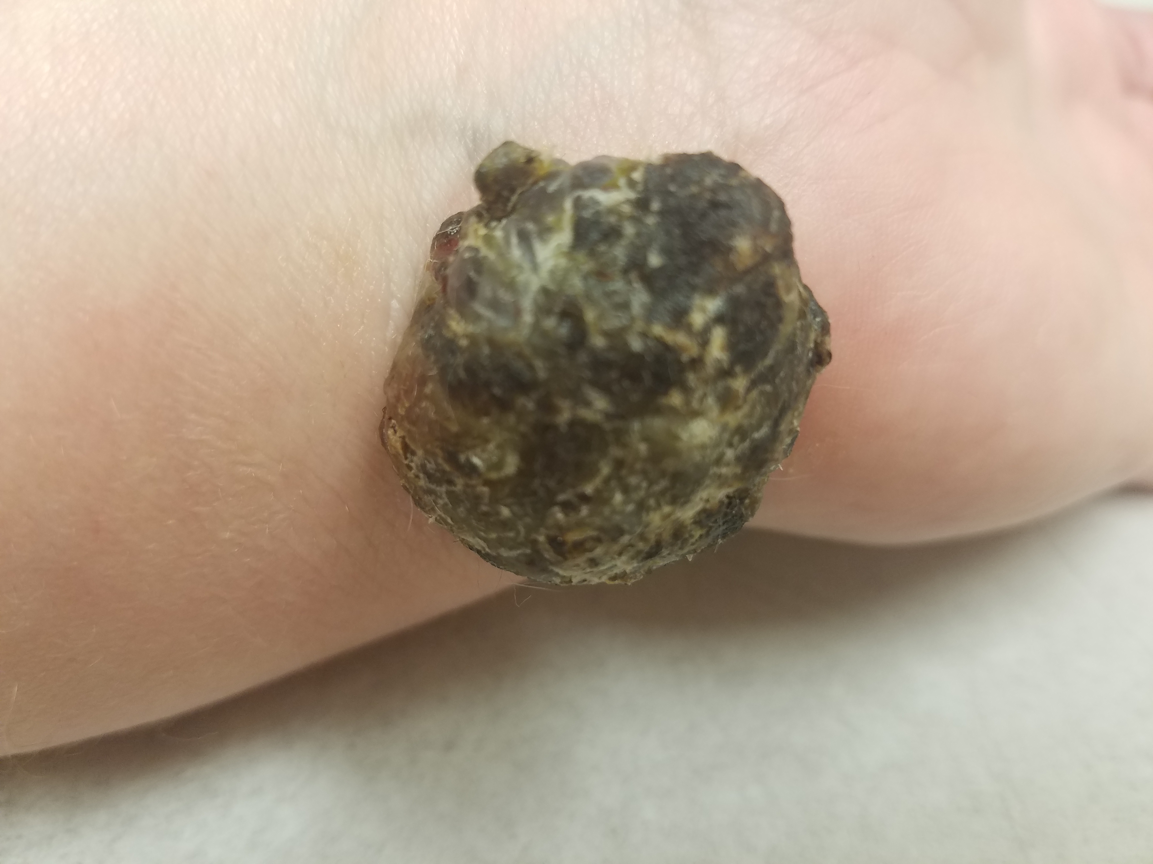
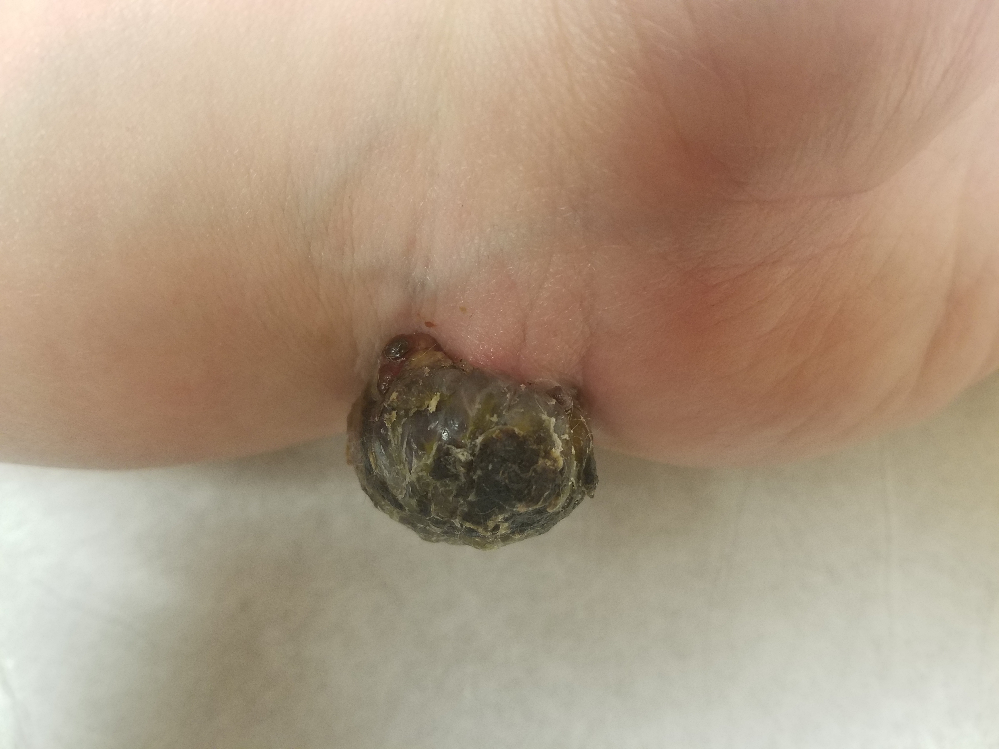
On physical examination, her vital signs were within normal limits except for a body mass index of 56 kg/m2. Examination of her left wrist showed a 4- to 5-cm, round, black-brown, pedunculated mass on the volar aspect of the left wrist (Figures). The mass was similar in size and shape to a ping-pong ball and was nontender. The rest of the physical examination findings were unremarkable.
Answer: Malignant Melanoma
DISCUSSION
Cutaneous malignant melanoma accounts for 3% to 5% percent of all skin cancers and is responsible for approximately 75% for all deaths from skin cancer.1 In the United States, it is the sixth most common cancer in both men and women.2 Cutaneous malignant melanoma can occur de novo or in an existing nevus; thus, close examination of all nevi on a patient is necessary.3,4 The ABCDE mnemonic is used for evaluating melanomas (Table 1).1
Table 1. The ABCDE Mnemonic | |
Asymmetry | One half of the lesion is different than the other half |
Border | Irregular or poorly defined border |
Color | Irregular color |
Diameter | Larger than 6 mm |
Evolving | Change in size, shape, or color |
The “ugly duckling” sign is a simple technique to aid in the detection of melanoma and is when a lesion stands out as different from the person’s other moles.1 The 4 major types of malignant melanoma are superficial spreading melanoma, nodular melanoma, lentigo maligna, and acral lentiginous melanoma. Other variants are amelanotic melanoma, subungual melanoma, spitzoid melanoma, desmoplastic melanoma, and pigment-synthesizing melanoma.
Dermoscopy can be an important tool in the diagnosis of melanoma.5 Saucerization shave, punch, and excisional biopsy are all acceptable techniques, provided they capture both the breadth and depth of the tumor. The decision about which technique to use depends on many factors including the clinician’s skill, pretest probability of melanoma, perception of lesion depth, lesion location, lesion size, equipment availability, patient healing, and cosmetic results.6
Commonly used markers for immunohistochemistry are S-100, MART-1, and HMB-45, and adjunctive diagnostic tests include comparative genomic hybridization, fluorescence in situ hybridization, gene expression profiling of tumors, and adhesive patch genomic analysis.7
Proper surgical management is critical in diagnosing, staging, and properly treating primary cutaneous melanoma. The Breslow depth of the lesion determines definitive surgical margins. Recommended surgical margins for melanoma based on Breslow depth are summarized in Table 2.1
Table 2. Recommended Surgical Margins for Melanoma Based on Breslow Depth | |
Breslow Depth | Margins |
In situ | 5 mm |
<2.0 mm | 1 cm |
>2.0 mm | 2 cm |
Treatment of lentigo maligna melanoma in particular is challenging. Standard excision with 5-mm to 1-cm margins have recurrence rates ranging from 8% to 50%.8 This is often approached as a staged procedure, where histologic clearance is confirmed prior to definitive reconstruction.8
Mohs micrographic surgery (MMS) involves stepwise tangential excision of specimen margins up to normal-appearing skin, followed by immediate microscopic examination of the entire surgical margin. Despite the advantage of MMS as a tissue-sparing procedure, controversy surrounds the use of frozen sections to identify malignant melanocytic cells. MMS is not an acceptable modality for invasive melanoma.8
In cases in which the Breslow depth is 1.0 mm or greater, the patient is referred for a sentinel lymph node biopsy. The stage of disease is determined by the TNM classification system, which considers the thickness of the tumor, the degree of nodal involvement, and the extent of metastatic spread to other areas of the body.1 Adjunctive immunotherapy with checkpoint inhibitors and targeted agents is recommended for patients with lymph node involvement. In patients with a large primary tumor who require extensive disfigurement, regional chemotherapy may be used to shrink the tumor prior to the procedure.1,9
REFERENCES:
- Shenenberger DW. Cutaneous malignant melanoma: a primary care perspective. Am Fam Physician. 2012;85(2):161-168.
- Siegel RL, Miller KD, Jemal A. Cancer statistics, 2018. CA Cancer J Clin. 2018;68(1):7-30.
- Skender-Kalnenas TM, English DR, Heenan PJ. Benign melanocytic lesions: risk markers or precursors of cutaneous melanoma? J Am Acad Dermatol. 1995;33(6):1000-1007.
- Goodson AG, Grossman D. Strategies for early melanoma detection: approaches to the patient with nevi. J Am Acad Dermatol. 2009;60(5):719-738. doi:10.1016/j.jaad.2008.10.065
- Vestergaard ME, Macaskill P, Holt PE, Menzies SW. Dermoscopy compared with naked eye examination for the diagnosis of primary melanoma: a meta-analysis of studies performed in a clinical setting. Br J Dermatol. 2008;159(3):669-676.
- Snyder A, West SE, Miles CM, Feldman SR. Obtaining an adequate specimen for the diagnosis of pigmented lesions. J Am Board Fam Med. 2015;28(4):523-525. doi:10.3122/jabfm.2015.04.150043
- Gerami P, Alsobrook JP II, Palmer TJ, Robin HS. Development of a novel noninvasive adhesive patch test for evaluation of pigmented lesions of the skin. J Am Acad Dermatol. 2014;71(2):237-244.
- Joyce KM. Surgical management of melanoma. In: Ward WH, Farma JM, eds. Cutaneous Melanoma: Etiology and Therapy. Brisbane, Australia: Codon Publications; 2017:chap 7. https://www.ncbi.nlm.nih.gov/books/NBK481850/. Accessed December 2, 2019.
- Higgins JC, Maher MH, Douglas MS. Diagnosing common benign skin tumors. Am Fam Physician. 2015;92(7):601-607.