
Peer Reviewed
Ranula in a 13-Year-Old Girl
Authors:
Nishit Harishbhai Patel, MD
Assistant Professor, Department of Pediatrics, Division of Pediatric Emergency Medicine, UT Southwestern Medical Center, Dallas, Texas
Shashidhar Marneni, MD
Assistant Professor, Department of Pediatrics, Division of Pediatric Emergency Medicine, UT Southwestern Medical Center, Dallas, Texas
Citation:
Patel NH, Marneni S. Ranula in a 13-year-old girl. Consultant. 2019;59(10):315-316.
A 13-year-old otherwise healthy girl presented to the emergency department (ED) with a 3-week history of progressive swelling of the right side of her neck and underneath the right side of her tongue. The swelling had been causing discomfort while eating. She denied any fever, vomiting, cough, changes in voice, difficulty breathing, oral injury, or oral surgery. Her immunization status was up to date.
On her physical examination, vital signs were within normal limits. A dome-shaped, bluish, translucent, firm, movable, and nontender swelling was present on the right-sided floor of the mouth (Figure 1). Also noted was an extension of the swelling to the right side of the girl’s neck, involving the submental and submandibular region (Figure 2), with no overlying redness or warmness of skin. Rest of her physical examinations were unremarkable.

Figure 1. Intraoral right-sided sublingual swelling (arrow).

Figure 2. Right-sided neck swelling (arrow).
A computed tomography (CT) scan of the neck with contrast showed a cystic lesion measuring approximately 4.5 cm in the largest dimension, with no significant peripheral enhancement, extending from the right sublingual space to the anterior submandibular space. Anteriorly, the lesion also crossed the midline to the left sublingual space (Figures 3-5). These imaging findings confirmed the clinically suspected diagnosis of ranula.

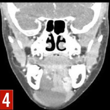

Figures 3-5. CT scan of the neck with contrast (axial view [3], coronal view, [4]; and sagittal view [5]) showed a 4.5-cm cystic lesion, with no significant peripheral enhancement, extending from the right sublingual space to the anterior submandibular space.
She was discharged home with a recommendation to follow up with an otorhinolaryngologist for further management. We were unable to follow up, because the girl had been visiting from out of town.
Discussion. A ranula is an uncommon, benign, and usually asymptomatic cystic lesion that arises from the obstruction or rupture of the excretory duct of the sublingual gland, which causes mucus extravasation into the surrounding tissue.1 A simple ranula is restricted to the oral cavity floor. A plunging ranula extravasates through the mylohyoid muscle, toward the cervical structures in the submandibular space, causing neck swelling.1,2
A ranula can present at any age. Oral trauma is considered a risk factor. The differential diagnosis includes dermoid cyst, epidermoid cyst, thyroglossal duct cyst, vascular malformations, soft tissue tumors, and salivary gland calculi or tumors. Imaging studies (ultrasonography, CT, or magnetic resonance imaging) can be helpful in determining the origin and extension of the lesion.3-5
The treatment modalities include observative or surgical methods (eg, excision of the lesion with or without excision of ipsilateral sublingual gland; marsupialization; cryosurgery; carbon dioxide laser excision). The prognosis depends on the treatment used.2,3,6
References:
- Bonet-Coloma C, Minguez-Martinez I, Aloy-Prósper A, Galán-Gil S, Peñarrocha-Diago M, Mínguez-Sanz JM. Pediatric oral ranula: clinical follow-up study of 57 cases. Med Oral Patol Oral Cir Bucal. 2011;16(2):e158-e162.
- Olojede ACO, Ogundana OM, Emeka CI, et al. Plunging ranula: surgical management of case series and the literature review. Clin Case Rep. 2017;6(1):109-114.
- Kokong D, Iduh A, Chukwu I, Mugu J, Nuhu S, Augustine S. Ranula: current concept of pathophysiologic basis and surgical management options. World J Surg. 2017;41(6):1476-1481.
- Jain P, Jain R, Morton RP, Ahmad Z. Plunging ranulas: high-resolution ultrasound for diagnosis and surgical management. Eur Radiol. 2010;20(6):1442-1449.
- Patel S, Bhatt AA. Imaging of the sublingual and submandibular spaces. Insights Imaging. 2018;9(3):391-401.
- Zhi K, Gao L, Ren W. What is new in management of pediatric ranula? Curr Opin Otolaryngol Head Neck Surg. 2014;22(6):525-529.