Peer Reviewed
How Would You Diagnose This Rash on a Young Woman?
Correct answer: E. Secondary syphilis
Secondary syphilis was diagnosed. Although several disease states can cause a rash in this distribution pattern, a high index of suspicion for syphilis should be maintained in patients who have had multiple sexual partners, a history of prior sexually transmitted infections, and in men who have sex with men. Data from the Centers for Disease Control and Prevention show that the rate of syphilis reached an all-time low in 2000 since reporting began in 1941.1 However, the rate has been increasing since 2000. From 2018 to 2019, there was an 11.2% increase in the number of reported syphilis cases.1 The rise in cases is believed to be related to technology and the increased use of online dating apps that have made it easier to meet partners and engage in casual sex.
Secondary syphilis has a highly variable appearance, but lesions often appear as pale pink or hyperpigmented macules that involve the palms, soles, and flexor surfaces of the extremities, as seen in our patient. Screening is conducted with a nontreponemal test such as the rapid plasma reagin test.2 Nontreponemal tests may return false-positive results in patients who have an autoimmune disease, HIV, pregnancy, or history of drug use.3 This occurs when the patient has another disease that produces antibodies similar to those associated with syphilis. Given that our patient had antiphospholipid syndrome, we conducted a fluorescent treponemal antibody absorption test to confirm the diagnosis. Results were positive, indicating that she had syphilis.
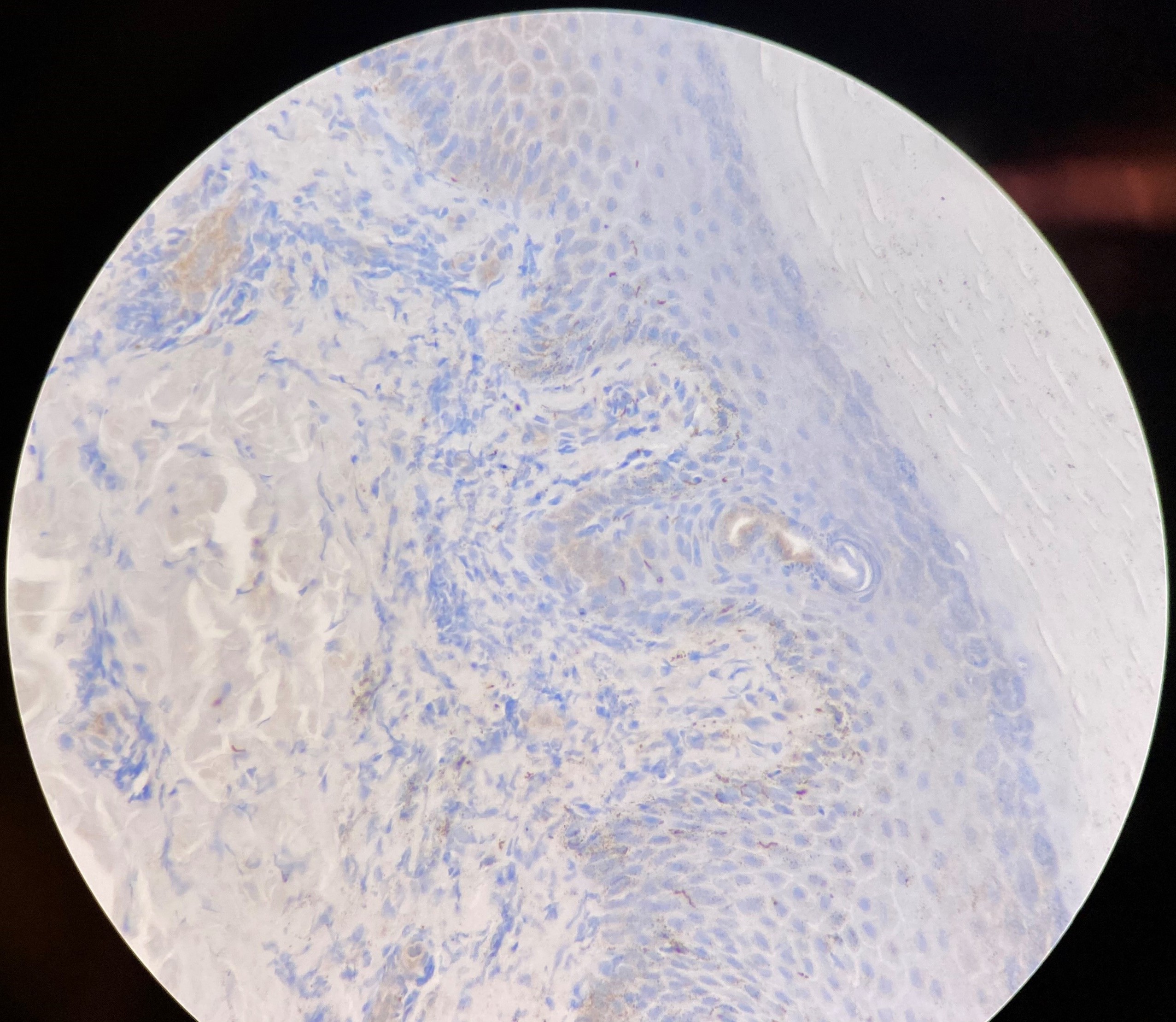
A punch biopsy was conducted at the initial visit, and perivascular plasma cells were seen, further confirming the diagnosis (Figure 3).4,5 A Treponema pallidum stain showed curly rods, representing spirochetes in syphilis (Figure 4).4,5 Spirochetal diseases often cause cutaneous or mucosal manifestations, affect multiple systems such as neurologic and cardiovascular, and have stages as well as latent periods.5 Dense plasma cell infiltrates and spirochetes can also be seen in Borrelia burgdorferi (Lyme disease).4 Other diseases in which spirochetes are seen include Leptospira interrogans (leptospirosis) and Treponema pallidum pertenue (Yaws). Our patient’s history, demographics, presentation, and laboratory results were most consistent Treponema pallidum.5

Figure 3. A hematoxylin and eosin stain taken from a punch biopsy of the patient’s left plantar surface showed numerous perivascular plasma cells (circle), which can be identified by an eccentric nucleus and a perinuclear clearing known as the Hof (arrow). The Hof is a lightly stained area proximal to the nucleus that represents the Golgi apparatus, which functions to package protein.
Figure 4. A Treponema pallidum immunostain showed spirochetes, which are elongated, tightly coiled bacteria with tapered ends. Motility occurs via periplasmic flagella or endoflagella.Treatment and management. Treatment for primary, secondary, and early latent syphilis (< 12 months) is with intramuscular penicillin G benzathine, 2.4 units, as a single dose. If the infection is latent (≥ 12 months) or duration is unknown, the treatment is with intramuscular penicillin G benzathine, 2.4 million units, weekly for 3 weeks.3
Discussion. Syphilis is often called “the Great Imitator” since it can present similarly to many other diseases. Erythema multiforme caused by an adverse drug reaction was a strong consideration in this case since the rash had appeared close to the time she had started warfarin. Erythema multiforme is an immune-mediated reaction that causes targetoid lesions associated with viral infections, Mycoplasma pneumoniae, and several medications.6 It can also occur in response to medications such as warfarin.7 Our patient was switched to apixaban 3 weeks prior to presentation at our clinic, and the rash had persisted, putting erythema multiforme lower on our differential diagnosis. Furthermore, the positive serology and histology findings were consistent with syphilis.
Cutaneous lupus erythematosus presents as papulosquamous erythematous plaques primarily on sun-exposed areas of the body such as the shoulders, extensor surface of the upper extremities, and neck but often spare the face.8 Patients will have a history of photosensitivity, and laboratory test results will often be positive for antinuclear antibodies and lupus anticoagulant.8 Our patient did not have those antibodies. In addition, the punch biopsy and clinical examination findings were not consistent with cutaneous lupus erythematosus. Results of a punch biopsy would have shown lymphocytic infiltrates along the dermal-epidermal junction and hyperkeratosis along with thickening of the basement membrane.9 Immunofluorescence would have shown immunoglobulin A, G, and M at the dermo-epidermal junction.9
Rocky Mountain spotted fever, which is caused by Rickettsia rickettsii, is carried by the Dermacentor variabilis tick.10 Patients would have headache, fever, and maculopapular rash that progresses to petechiae.10 The rash starts on the palms and soles and spreads centrally to the trunk, sparing the face.10 Clinicians should maintain a high index of suspicion among patients who have recently spent a great deal of time outdoors, especially in the summer months in the southeastern and south-central parts of the United States.10 Our patient had not traveled recently, and she did not spend a great deal of time outdoors. In addition, she did not have the other symptoms such as headache or fever. For these reasons, Rocky Mountain spotted fever was lower on the differential diagnosis.
Lichen planus is an immunologically mediated skin disorder most commonly affecting middle-aged adults.11 It involves the skin and mucous membranes of the mouth, nails, and external genitalia.11 It is often remembered as the 6 Ps: planar (flat-topped), purple, pruritic, polygonal, plaques, and papules (2-5 mm in size).11 Lichen planus affects the flexural surfaces (most commonly the wrists), buccal mucosa, and external genitalia.11 Wickham striae, which are white lacy streaks, often appear on the surface of the lesions, especially on the tongue and buccal mucosa.12 Lichen planus has been associated with hepatitis C virus, but no specific causal relationship has been established between the two.12 Screening for hepatitis C virus should be considered in patients with lichen planus. Hematoxylin and eosin staining will classically show civatte bodies, which are round eosinophilic masses in the epithelial layer.13
Patient outcome. Our patient was treated with 3 weekly doses of intramuscular penicillin G, as her history was not clear in terms of the duration of infection. Therefore, after a discussion with the patient, we opted to err on the side of caution and minimize her risk. She did receive all 3 doses on subsequent visits. Moreover, her rash had completely resolved by the third follow-up visit. Results from repeat rapid plasma reagin titers are still pending, but the patient was advised to follow up in 6 months from the original tests to repeat the titers.
References
1. National Overview - Sexually Transmitted Disease Surveillance, 2019. Syphilis. Centers for Disease Control and Prevention. Reviewed April 13, 2021. Accessed March 17, 2021. https://www.cdc.gov/std/statistics/2019/overview.htm#Syphilis
2. Cantor AG, Pappas M, Daeges M, Nelson HD. Screening for syphilis: updated evidence report and systematic review for the US Preventive Services Task Force. JAMA. 2016;315(21):2328-2337. https://doi.org/10.1001/jama.2016.4114
3. Workowski KA, Bachmann LH, Chan PA, et al. Sexually transmitted infections treatment guidelines, 2021. MMWR Recomm Rep. 2021;70(4):1-187. https://doi.org/10.15585/mmwr.rr7004a1
4. Carlson JA, Dabiri G, Cribier B, Sell S. The immunopathobiology of syphilis: the manifestations and course of syphilis are determined by the level of delayed-type hypersensitivity. Am J Dermatopathol. 2011;33(5):433-460. https://doi.org/10.1097/dad.0b013e3181e8b587
5. Giacani L, Lukehart SA. The endemic treponematoses. Clin Microbiol Rev. 2014;27(1):89-115. https://doi.org/10.1128/cmr.00070-13
6. Trayes KP, Love G, Studdiford JS. Erythema multiforme: recognition and management. Am Fam Physician. 2019;100(2):82-88. https://www.aafp.org/afp/2019/0715/p82.html
7. Svensson CK, Cowen EW, Gaspari AA. Cutaneous drug reactions. Pharmacol Rev. 2001;53(3):357-379. https://pharmrev.aspetjournals.org/content/53/3/357
8. Walling HW, Sontheimer RD. Cutaneous lupus erythematosus: issues in diagnosis and treatment. Am J Clin Dermatol. 2009;10(6):365-381. https://doi.org/10.2165/11310780-000000000-00000
9. Cheng H, Lamont D, Emanuel P. Discoid lupus erythematosus pathology. DermNet NZ. Published 2014. Accessed March 17, 2021. https://dermnetnz.org/topics/discoid-lupus-erythematosus-pathology/
10. Biggs HM, Behravesh CB, Bradley KK, et al. Diagnosis and management of tickborne rickettsial diseases: Rocky Mountain spotted fever and other spotted fever group rickettsioses, ehrlichioses, and anaplasmosis - United States. MMWR Recomm Rep. 2016;65(2):1-44. https://doi.org/10.15585/mmwr.rr6502a1
11. Gorouhi F, Davari P, Fazel N. Cutaneous and mucosal lichen planus: a comprehensive review of clinical subtypes, risk factors, diagnosis, and prognosis. ScientificWorldJournal. 2014;2014:742826. https://doi.org/10.1155/2014/742826
12. Lodi G, Pellicano R, Carrozzo M. Hepatitis C virus infection and lichen planus: a systematic review with meta-analysis. Oral Dis. 2010;16(7):601-612. https://doi.org/10.1111/j.1601-0825.2010.01670.x
13. Pranay T, Kumar AS, Chhabra S. Civatte bodies: a diagnostic clue. Indian J Dermatol. 2013;58(4):327. https://doi.org/10.4103/0019-5154.113974