
Peer Reviewed
A 13-Year-Old Girl With a Bilateral Axillary Rash
AUTHORS:
Courtney Humphrey, MD • Gardia Germinal, MD
AFFILIATIONS:
St. Luke’s Family Medicine Residency, Bethlehem, Pennsylvania
CITATION:
Humphrey C, Germinal G. A 13-year-old girl with a bilateral axillary rash. Consultant. 2021;61(5):e9-e11. doi:10.25270/con.2020.09.00007
Received April 8, 2020. Accepted June 16, 2020. Published online September 9, 2020.
DISCLOSURES:
The authors report no relevant financial relationships.
CORRESPONDENCE:
Courtney Humphrey, MD, Clinical Faculty, St. Luke’s Family Medicine Residency Program, 2830 Easton Ave, Bethlehem, PA 18017 (courtney.humphrey@sluhn.org)
A 13-year-old girl presented to the clinic with hair loss and a rash in the axillae for 4 months. The lesions initially had appeared as erythematous and coin-shaped and then had evolved into hyperpigmented plaques. At this initial presentation, she was diagnosed with tinea capitis and tinea corporis and was prescribed a 4-week course of griseofulvin, which led to resolution of the lesions.
One month after the initial resolution of the rash, however, the hyperpigmented plaques reappeared in the girl’s the armpits, prompting another visit. At this visit, she was prescribed nystatin topical powder, the application of which led no improvement. The rash became pruritic, and she presented again to the office 1 month later.
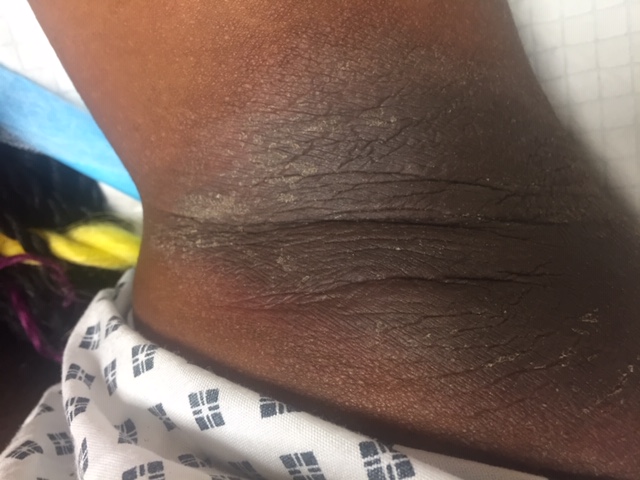
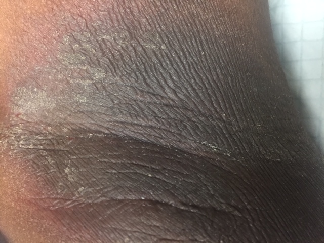
Physical examination showed a well-demarcated, hyperpigmented, lichenified rash with multiple excoriations and fissures in the axillae (Figures). The skin in the area was warm and mildly tender to the touch. There was no evidence of fungal infection on Wood lamp examination, and biopsy revealed psoriasiform epidermal hyperplasia with parakeratosis and mild spongiosis. Recent screening test results for diabetes were negative.


Figure. A well-demarcated, hyperpigmented, lichenified rash with multiple excoriations and fissures in the axillae of a 13-year-old girl.
Answer: Spongiotic Psoriasiform Dermatitis
Spongiotic psoriasiform dermatitis is a chronic inflammatory skin disorder that primarily affects children but can also affect adults. Typical locations include the chest, abdomen, and buttocks, but spongiotic psoriasiform dermatitis can affect areas that are atypical for eczema. This condition commonly manifests initially with severe pruritus, and afterwards, the rash appears. The rash at first appears red and then darkens to a dark brown color. If left untreated, spongiotic psoriasiform dermatitis can develop into weeping welts and later can become rough and crusty appearing.
Testing is not necessary unless the patient is not responding to therapy. In this case, a biopsy is the best diagnostic modality.
Topical agents are the preferred treatment for this condition. Menthol-based formulations are the first line agents and are applied directly onto the skin.1 Menthol serves as a topical analgesic with antipruritic properties that can enhance transdermal drug delivery. If menthol-based formulations are ineffective, topical corticosteroids may be added. Potency should be based on location and severity of the lesions. Low-potency corticosteroids (such as desonide, gel, cream, ointment, or foam, 0.05%, or fluocinolone acetonide cream, 0.01%, twice daily) can be used for mild dermatitis.1 Medium-potency corticosteroids (such as betamethasone valerate cream or lotion, 0.1%, or fluticasone propionate cream, 0.05%, twice daily) can be used for moderate dermatitis. Creams are preferred for large and subacute areas while ointments are preferred for dry and lichenified areas; lotions may help to cool or dry inflamed and oozing lesions.1
DIFFERENTIAL DIAGNOSIS
Psoriasis is a chronic inflammatory skin disorder that affects the extensor surfaces such as the knees and elbows. The rash is raised with red scaly patches that are well demarcated. The condition is associated with atherosclerotic disease, diabetes, hypertension, metabolic syndrome, and obesity.2,3 The Koebner phenomenon (development of disease in areas of skin trauma) and Auspitz sign (pinpoint bleeding after removal of scale) are the classic clinical findings.4 A biopsy may show epidermal hyperplasia, parakeratosis, neutrophils in the stratum corneum, or thinned granular layer of epidermis.5
Erythrasma is a superficial infection of the skin caused by Corynebacterium minutissimum, a gram-positive, non–spore-forming bacillus.6 It presents as a macerated, scaly, and erythematous to thin brown rash in the intertriginous and interdigital areas.6 The bacteria proliferate in the stratum corneum of moist environments such as the toe webs, groin, and axillae.6,7 Wood lamp examination reveals coral-red fluorescence. Skin scraping with a potassium hydroxide preparation is another diagnostic test in patients with interdigital involvement.6 Topical clindamycin and erythromycin are first-line treatments, while topical antifungals such as miconazole, oxiconazole, or econazole are an alternative.8,9
Seborrheic dermatitis is a chronic relapsing dermatitis characterized by well-demarcated, erythematous plaques with greasy-looking, yellowish scales on areas rich in sebaceous glands such as the scalp, face, and trunk. It is commonly seen in healthy individuals but may affect persons in immunocompromised states, those on neuroleptic medications, or those with neurologic disorders such as Parkinson disease.10 Although not a disease of the sebaceous gland, it does play a permissive role by creating a favorable environment for growth. The diagnosis is made clinically but may require biopsy when unclear. Treatment is dependent on the number of areas involved and previous treatments. Topical antifungal and anti-inflammatory agents are typically used, especially for the face, intertriginous areas, scalp, and trunk. Immunocompromised patients require oral agents such as Itraconazole, ketoconazole, fluconazole, or terbinafine.11
Acanthosis nigricans is a common skin condition with velvety, hyperpigmented plaques commonly found in the neck and axillae. It is associated with insulin resistance such as diabetes or obesity and can manifest in certain malignancies.12 Hyperkeratosis, and epidermal papillomatosis are the major histologic features.13 Treatment of the underlying conditions is the preferred treatment; if this is not achievable, topical retinoid and topical vitamin D analogue therapy may be beneficial.14,15
OUTCOME OF THE CASE
After biopsy results confirmed the diagnosis of spongiotic psoriasiform dermatitis, the patient was started on topical corticosteroids and was instructed to apply it to the axillae twice daily for 7 days. At a well-child visit 3 months later, the patient reported complete resolution of the rash.
REFERENCES:
- Tadicherla S, Ross K, Shenefelt PD, Fenske NA. Topical corticosteroids in dermatology. J Drugs Dermatol. 2009;8(12):1093-1105.
- Armstrong AW, Harskamp CT, Armstrong EJ. The association between psoriasis and obesity: a systematic review and meta-analysis of observational studies. Nutr Diabetes. 2012;2(12):e54. doi:10.1038/nutd.2012.26
- Rodríguez-Zúñiga MJM, García-Perdomo HA. Systematic review and meta-analysis of the association between psoriasis and metabolic syndrome. J Am Acad Dermatol. 2017;77(4):657-666.e8. doi:10.1016/j.jaad.2017.04.1133
- Bernhard JD. Auspitz sign is not sensitive or specific for psoriasis. J Am Acad Dermatol. 1990;22(6 pt 1):1079-1081. doi:10.1016/0190-9622(90)70155-b
- Grover C, Reddy BSN, Uma Chaturvedi K. Diagnosis of nail psoriasis: importance of biopsy and histopathology. Br J Dermatol. 2005;153(6):1153-1158. doi:10.1111/j.1365-2133.2005.06862.x
- Svejgaard E, Christophersen J, Jelsdorf HM. Tinea pedis and erythrasma in Danish recruits: clinical signs, prevalence, incidence, and correlation to atopy. J Am Acad Dermatol. 1986;14(6):993-999. doi:10.1016/s0190-9622(86)70122-9
- Inci M, Serarslan G, Ozer B, et al. The prevalence of interdigital erythrasma in southern region of Turkey. J Eur Acad Dermatol Venereol. 2012;26(11):1372-1376. doi:10.1111/j.1468-3083.2011.04293.x
- Holdiness MR. Management of cutaneous erythrasma. Drugs. 2002;62(8):1131-1141. doi:10.2165/00003495-200262080-00002
- Cochran RJ, Rosen T, Landers T. Topical treatment for erythrasma. Int J Dermatol. 1981;20(8):562-564. doi:10.1111/j.1365-4362.1981.tb02030.x
- Kohn SR, Pochi PE, Strauss JS, Sax DS, Feldman RG, Timberlake WH. Sebaceous gland secretion in Parkinson’s disease during L-dopa treatment. J Invest Dermatol. 1973;60(3):134-136. doi:10.1111/1523-1747.ep12682040
- Apasrawirote W, Udompataikul M, Rattanamongkolgul S. Topical antifungal agents for seborrheic dermatitis: systematic review and meta-analysis. J Med Assoc Thai. 2011;94(6):756-760.
- Habif TP. Acanthosis nigricans. In: Habif TP. Clinical Dermatology: A Color Guide to Diagnosis and Therapy. 5th ed. Mosby Elsevier;2010:978-979.
- Weedon D. Miscellaneous conditions. In: Weedon D. Weedon’s Skin Pathology. 3rd ed. Churchill Livingston Elsevier; 2010:chap 19.
- Adigun CG, Pandya AG. Improvement of idiopathic acanthosis nigricans with a triple combination depigmenting cream. J Eur Acad Dermatol Venereol. 2009;23(4):486-487. doi:10.1111/j.1468-3083.2008.02931.x
- Blobstein SH. Topical therapy with tretinoin and ammonium lactate for acanthosis nigricans associated with obesity. Cutis. 2003;71(1):33-34.