
Peer Reviewed
Pustular Scab on an Elderly Woman's Head
Answer: C. Erosive pustular dermatosis
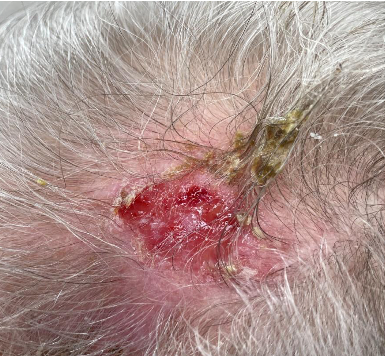
Given the patient's age, years of sun exposure, and recent history of electrodessication and curettage to the scalp, a diagnosis of erosive pustular dermatosis of the scalp (EPDS) was made. The patient was informed that the removal of hypertrophic actinic keratosis by electrodessication and curettage about 1 month earlier had likely triggered the development of EPDS. After the crust was removed from her scalp (Figure 2), she was given topical clobetasol ointment to apply to the lesion twice daily and instructed to make a follow-up appointment in 2 weeks for further evaluation. The patient was also counseled on the importance of proper sun protection of her scalp, as cutaneous atrophy secondary to chronic sun exposure increases the risk and progression of EPDS.1

Figure 2. Removal of crust reveals an atrophic base with prominent erythema around previous electrodessication and curettage on the patient's scalp.
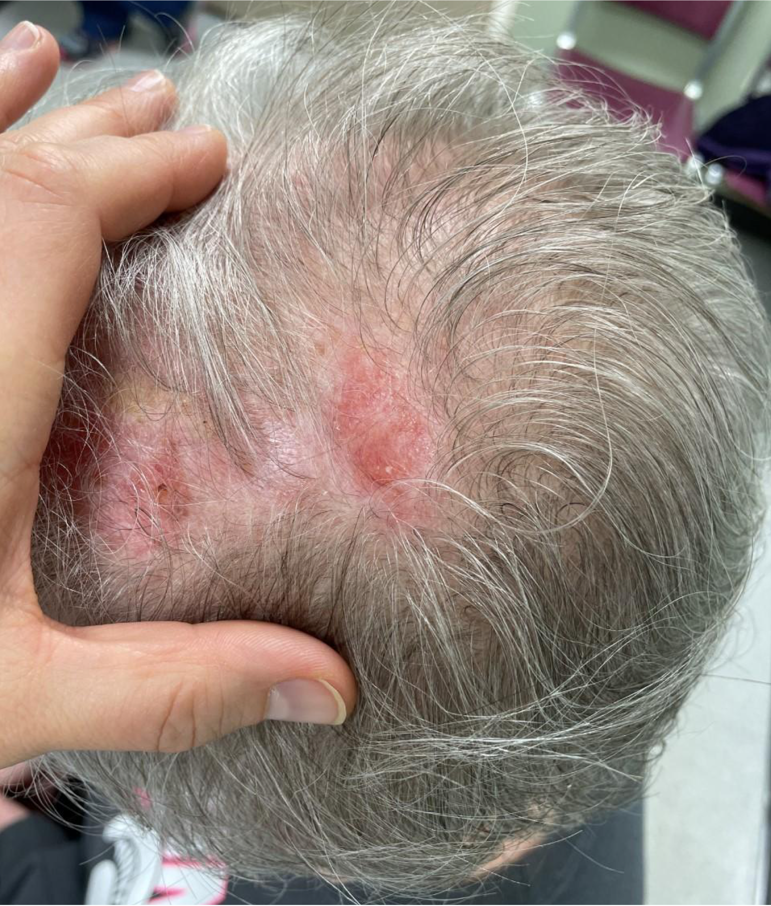
Patient outcome. Two weeks later at the follow-up visit, the patient reported using the topical clobetasol ointment as prescribed and noted a significant improvement in her EPDS (Figure 3). On physical examination, we noted significant atrophy and alopecia where the crust had originally been. Minimal erythema was present, but other exudates or pus were absent. She was instructed to discontinue the clobetasol ointment to prevent further steroid-induced atrophy and to continue sun protection measures. Because of the increased risk of skin malignancy (eg, squamous cell carcinoma) developing on scar tissue in EPDS lesions,2 the patient was advised to receive annual skin exams of her scalp and full body.
Figure 3. Alopecia and atrophy with mild erythema remain after 2 weeks of clobetasol ointment applied twice daily to the patient's EPDS lesion.
Discussion. Also known as erosive pustulosis of the scalp, EPDS is an insidious and chronic inflammatory skin condition characterized by yellow-brown crusted lesions, pustules, or erosion that result in scarring alopecia. Initially described by Burton3 in 1977and Pye and colleagues4 in 1979,EPDS has a predilection for sun-damaged skin among older adults. This rare condition is difficult to diagnose because it often mimics common conditions of sun-damaged skin (eg, actinic keratosis) and has nonspecific clinical, dermoscopic, and histopathologic features.5
As seen in our patient, EPDS may develop secondary to skin trauma (eg, electrodessication and curettage, cosmetic procedures, sun or thermal burns, chemotherapy, and surgeries).6 The pathophysiology of EPDS is poorly understood, however, it typically develops after cutaneous atrophy and actinic damage. Trauma to the damaged skin can be a triggering factor; as in our case, the patient's treatment of actinic keratoses was associated with the development of erosive pustulosis. High potency steroids (eg, clobetasol) and calcineurin inhibitors (eg, tacrolimus) are first-line therapy for EDPS.7,8
Dissecting cellulitis of the scalp is a chronic inflammatory disease and part of the follicular occlusion triad. Like EPDS, dissecting cellulitis can result in scarring alopecia. However, unlike EPDS, it is caused by inflammation at the bulb of the hair follicle, creating pustules that form into sinus tracts. Dissecting cellulitis is often refractory to treatment and requires surgical intervention.9
Ecthyma is an infection deep within the skin most often caused by streptococcus bacteria.10 The infection often blisters and leaves a crusted ulcer that may present similarly to the pustular exudate seen in our patient.10 Treatment involves antibiotics and warm compresses to remove the crusted ulcers. Ecthyma sometimes results in scarring.10
Another condition that results in scarring alopecia is folliculitis decalvans, a condition characterized by painful follicular exudates and neutrophilic infiltrates often presenting among immunocompromised adults.11 Treatment includes antibiotics for clearing the bacterial infection and anti-inflammatory medications.11 Our patient had no evidence of infection, which helped us rule out ecthyma and folliculitis decalvans.
Conclusion. With an examination of our patient and consideration of differential diagnoses, we were able to correctly diagnose and treat her condition. However, this case exemplifies that prevention is superior to treatment regarding the sun's harmful effects. Erosive pustulosis of the scalp is one of the many reasons to educate patients on the importance of sun protection. Treatment of sun damage is not always simple and can lead to complications, including erosive pustulosis of the scalp from actinic keratosis removal.
- Karanfilian KM, Wassef C. Erosive pustular dermatosis of the scalp: causes and treatments. Int J Dermatol. 2021;60(1):25-32. doi:10.1111/ijd.14955
- Wilk M, Zelger BG, Hauser U, Höpfl R, Zelger B. Erosive pustular dermatosis of the scalp: reappraisal of an underrecognized entity. J Dtsch Dermatol Ges. 2018;16(1):15-19. doi:10.1111/ddg.13387
- Burton JL. Case for diagnosis. Pustular dermatosis of scalp. Br J Dermatol. 1977;97 Suppl 15:67-69. doi:10.1111/j.1365-2133.1977.tb14339.x
- Pye RJ, Peachey RD, Burton JL. Erosive pustular dermatosis of the scalp. Br J Dermatol. 1979;100(5):559-566. doi:10.1111/j.1365-2133.1979.tb05581.x
- Piccolo V, Russo T, Bianco S, Ronchi A, Alfano R, Argenziano G. Erosive pustular dermatosis of the scalp: why do we miss it? Dermatology. 2019;235(5):390-395. doi:10.1159/000500909
- Robles DT. Erosive pustulosis of the scalp. Medscape. Published November 8, 2021. Accessed December 29, 2021. https://emedicine.medscape.com/article/1404360-overview
- Laffitte E, Kaya G, Piguet V, Saurat J. Erosive pustular dermatosis of the scalp: treatment with topical tacrolimus. Arch Dermatol. 2003;139(6):712-714. doi:10.1001/archderm.139.6.712
- Starace M, Iorizzo M, Trüeb RM, et al. Erosive pustular dermatosis of the scalp: a multicentre study. J Eur Acad Dermatol Venereol. 2020;34(6):1348-1354. doi:10.1111/jdv.16211
- Cuellar TA, Roh DS, Sampson CE. Dissecting cellulitis of the scalp: a review and case studies of surgical reconstruction. Plast Reconstr Surg Glob Open. 2020;8(8):e3015. doi:10.1097/GOX.0000000000003015
- Ecthyma. MedlinePlus. Updated May 2, 2021. Accessed January 5, 2022. https://medlineplus.gov/ency/article/000864.htm
- Otberg N, Kang H, Alzolibani AA, Shapiro J. Folliculitis decalvans. Dermatol Ther. 2008;21(4):238-244. doi:10.1111/j.1529-8019.2008.00204.x