
A 5-Year-Old presents with "itchy eczema"

A 5-year-old girl with atopic dermati- tis is brought to your office with a 4-day history of what the mother describes as “itchy eczema” on her legs.
What are your thoughts about the cause of the rash?
A. Atopic dermatitis flare.
B. Impetigo.
C. Herpes simplex.
D. Molluscum contagiosum.
E. Candidiasis.
Answer
The sudden onset of this oozing, crusted eruption suggested more than just a flare of eczema. The patient’s persistent scratching had caused a secondary staphylococcal eruption, impetigo, B.
Herpes simplex occurs in persons with atopy; however, the eruption usually is vesicular and painful. Candidiasis can mimic impetigo, but it is seen much less frequently. Molluscum contagiosum features umbilicated papules.
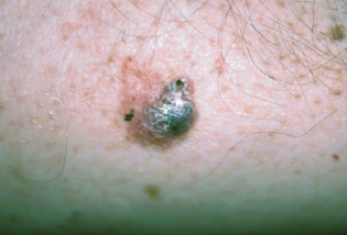
Twenty years after an asymptomatic growth erupted on his shin, a 77-year-old man seeks medical evaluation of the slowly enlarging lesion.
What do you suspect?
A. Seborrheic keratosis.
B. Basal cell carcinoma.
C. Melanoma.
D. Venous angioma.
E. Blue nevus.
Answer
The lesion’s pigmentation and general appearance suggested a possible skin cancer. A punch biopsy confirmed the diagnosis of basal cell carcinoma, B. Because of the possibility of melanoma, the biopsy specimen needs to include the base of the tumor.
A bluish lesion is not typical of seborrheic keratoses. The appearance of venous angiomas can be deceptive, but they usually look more vascular than this lesion. Blue nevi may be nodular; rarely, they undergo malignant degeneration.
This basal cell carcinoma—the only such lesion on this patient—was surgically removed without complications.
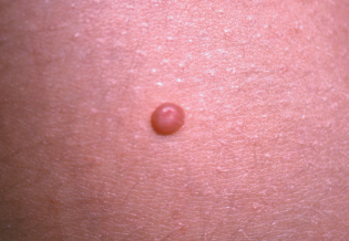
The parents of a 6-year-old boy are concerned about the asymptomatic bumps that have developed on their son’s abdomen during the last few months.
Do you recognize this lesion?
A. Molluscum contagiosum.
B. Wart.
C. Intradermal nevus.
D. Milium.
E. Keratosis pilaris.
Answer
This child had contracted molluscum contagiosum, A, a viral infection that resembles warts. The papules’ umbilicated center is the clinical clue to the diagnosis.
Warts have a rough, or verrucous, surface. Intra- dermal nevi lack a depressed center and do not usually develop on the abdomen of children. A milium appears as a deep whitehead-type comedo and does not often occur on the abdomen. The extremities and cheeks are common sites of keratosis pilaris, follicular plugs that affect persons with atopy.
Eight months earlier, a short, tapered dose of prednisone resolved pityriasis rosea in this patient. Now, the 14-year-old boy presents with a similar, mildly pruritic rash on his trunk. He is otherwise healthy, having fully recovered from an upper respiratory tract infection 3 or 4 weeks ago. Ten days ago, he returned from a class camping trip. The patient takes no medication.
Which of the following would you consider?
A. Recurrence of pityriasis rosea.
B. Guttate psoriasis.
C. Tinea corporis.
D. Reaction to an insect bite.
E. Contact dermatitis.
Answer
Pityriasis rosea, A, can occur more than once, and often develops after a recent upper respiratory tract infection. The diagnosis 8 months earlier was correct.
Guttate psoriasis is usually seen on the legs and does not respond to a tapering course of prednisone. Tinea corporis in an otherwise healthy person is not as extensive
as this patient’s eruption. Insect bites and contact dermatitis typically are not flat, scaly patches that are confined to the trunk.
A 15-year-old girl seeks treatment of asymptomatic white streaks that appeared on her trunk and thighs a few months earlier. The patient is otherwise healthy, takes no medica- tions, and has not had any signifi- cant weight changes in the past year.
What is the likely diagnosis?
A. Tinea versicolor.
B. Pityriasis alba.
C. Vitiligo.
D. Striae.
E. Scars from physical abuse.
Answer
Pubertal striae, D, developed as a result of rapid growth. These lesions are usually found on the back and around the breasts and thighs of adolescent girls and young women, and on the upper arms and back of adolescent boys and young men.
Tinea versicolor features discrete hypopigmented or hyperpigmented macules that typically appear on the trunk. Pityriasis alba manifests in persons with atopy as ill defined, hypopigmented patches. The patches of vitiligo are depigmented. Traumatic injuries generally have a linear or irregular scarring pattern that reflects the source of injury.