
Peer Reviewed
Eczema Coxsackium
Authors:
Kelley Ward, MD
Department of Pediatrics, University of Florida, Gainesville, Florida
Jeremy Smith
Medical Student, College of Medicine, University of Florida, Gainesville, Florida
Avni Bhatt, PhD
Department of Pediatrics, University of Florida, Gainesville, Florida
Mary Katherine Siebenaler, MD
Department of Pediatrics, Northwestern University Feinberg School of Medicine, and Ann & Robert H. Lurie Children’s Hospital of Chicago, Illinois
Citation:
Ward K, Smith J, Bhatt A, Siebenaler MK. Eczema coxsackium [published online September 11, 2019]. Consultant360.
A 13-month-old boy with a history of atopic dermatitis presented to the emergency department with a 4-day history of a rash preceded by a low-grade fever, rhinorrhea, and congestion. The rash had begun on his lower extremities, then had spread to his arms and face.
On physical examination, the rash was extensive and appeared confluent, erythematous, and maculopapular with vesicular lesions and was concentrated on his palms, face, and extremities, particularly in his areas of atopic dermatitis (Figures 1 and 2).

Figure 1. A vesiculopapular rash clustered on areas of prior atopic dermatitis. Perioral erosions are also present.
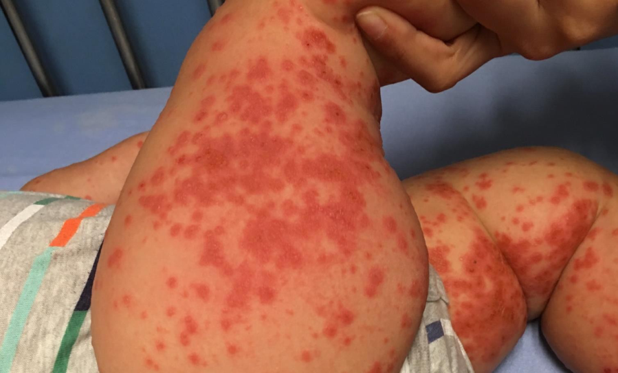
Figure 2. The vesiculopapular rash was most pronounced on the flexor surfaces of the lower extremities.
His posterior oropharynx had vesicles surrounded by halos of erythema, and perioral facial erosions were present. There were no lesions on his trunk. One day prior to admission, he had been prescribed cephalexin for the rash, for which he had taken as a single dose. Antibiotics were not continued during admission.
Upon presentation, the differential diagnosis included eczema coxsackium, eczema herpeticum, Giannotti Crosti syndrome, and bullous impetigo. An adverse drug reaction was considered, although it was deemed unlikely given the distribution of the rash and that antibiotics had been initiated after the onset of the rash.
The patient was observed overnight on intravenous fluids, with symptomatic relief for pruritis with calamine lotion and diphenhydramine. He was discharged on empirical treatment with acyclovir while awaiting the results of herpes simplex virus (HSV) polymerase chain reaction (PCR) testing of an unroofed vesicle. The PCR results returned negative for HSV-1 and HSV-2, making eczema herpeticum unlikely; the family was notified, and acyclovir was stopped.
The morphology of the rash with associated herpangina and facial erosions was highly suggestive of eczema coxsackium. Topical mupirocin was continued under his nares, given honey crusting lesions likely consistent with a secondary impetigo. A medium-potency topical corticosteroid (mometasone furoate cream, 0.1%) was initiated for eczematous areas. Close outpatient follow-up was arranged with the patient’s primary care pediatrician and a pediatric dermatologist.
DISCUSSION
Eczema coxsackium is an atypical viral exanthem generally caused by coxsackievirus 6.1 Classic hand-foot-and-mouth disease (HFMD) is a more commonly recognized exanthem, is most often associated with coxsackievirus A16 or enterovirus 71, and differs from eczema coxsackium in its appearance. The rash of HFMD often consists of vesicles or ulcerations of the buccal mucosa or posterior oropharynx.1 The skin manifestations of eczema coxsackium are variable and can include widespread eruption of vesicles, bullae, and erosions in a broader distribution than one would expect from HFMD, and it tends to be most prominent in areas of atopic dermatitis.
The lesions of eczema coxsackium may also appear on the cheeks, buttocks, and extensor surfaces, which can mimic the distribution of Giannotti Crosti syndrome, sparing the trunk, as in our patient’s case. Facial erosions and vesicles have also been described,2 as was also the case in our patient. Acral sites may show nonspecific nonblanching lesions consistent with petechiae or purpura.1,2
Associated symptoms include those of other enterovirus infections, such as fever, myalgia, gastroenteritis, meningitis, myocarditis, pancreatitis, and paralysis. A benign delayed manifestation can be nail changes including Beau lines or onychomadesis in addition to desquamation of the palms and soles.1 Our patient’s viral symptoms included malaise, upper respiratory tract symptoms, and fever.
The diagnosis of eczema coxsackium is made clinically, but if the diagnosis remains unclear, fluid from an unroofed vesical can be sent for PCR testing for confirmation of coxsackievirus infection.2 If eczema herpeticum is suspected, timely HSV PCR testing of the vesicular fluid is often helpful to guide acyclovir administration. Treatment involves supportive care, including pain control and hydration support, but wet wrapping and topical corticosteroid use have also been reported.3 The child should be observed for secondary bacterial infections, which are uncommon.1
This self-limited viral illness has an excellent prognosis, with lesions resolving without skin scarring, and it does not require long-term follow-up.
REFERENCES:
- Ventarola D, Bordone L, Silverberg N. Update on hand-foot-and-mouth disease. Clin Dermatol. 2015;33(3):340-346.
- Mathes EF, Oza V, Frieden IJ, et al. “Eczema coxsackium” and unusual cutaneous findings in an enterovirus outbreak. Pediatrics. 2013;132(1):e149-e157.
- Johnson VK, Hayman JL, McCarthy CA, Cardona ID. Successful treatment of eczema coxsackium with wet wrap therapy and low-dose topical corticosteroid. J Allergy Clin Immunol Pract. 2014;2(6):803-804.